. 2010 Sep;1(4):223-231.
doi: 10.1007/s13244-010-0035-6. Epub 2010 Jul 30.

Kawasaki disease: a review with emphasis on cardiovascular complications

Kawasaki disease: a review with emphasis on cardiovascular complications

Ricardo Duarte et al. Insights Imaging. 2010 Sep.

Abstract

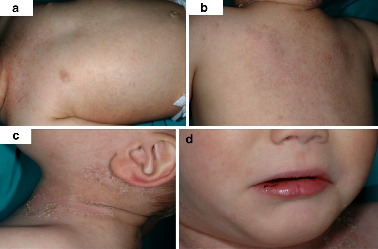
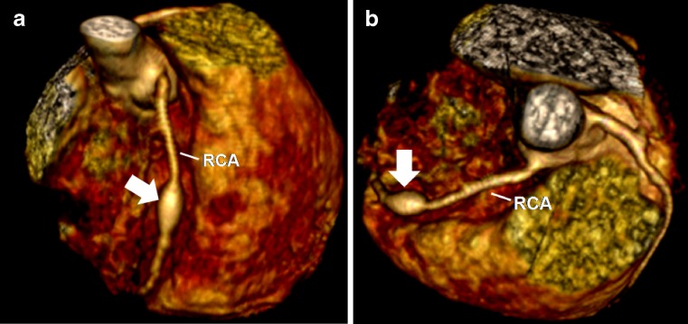
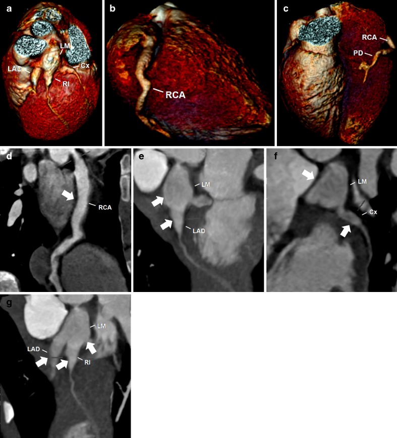
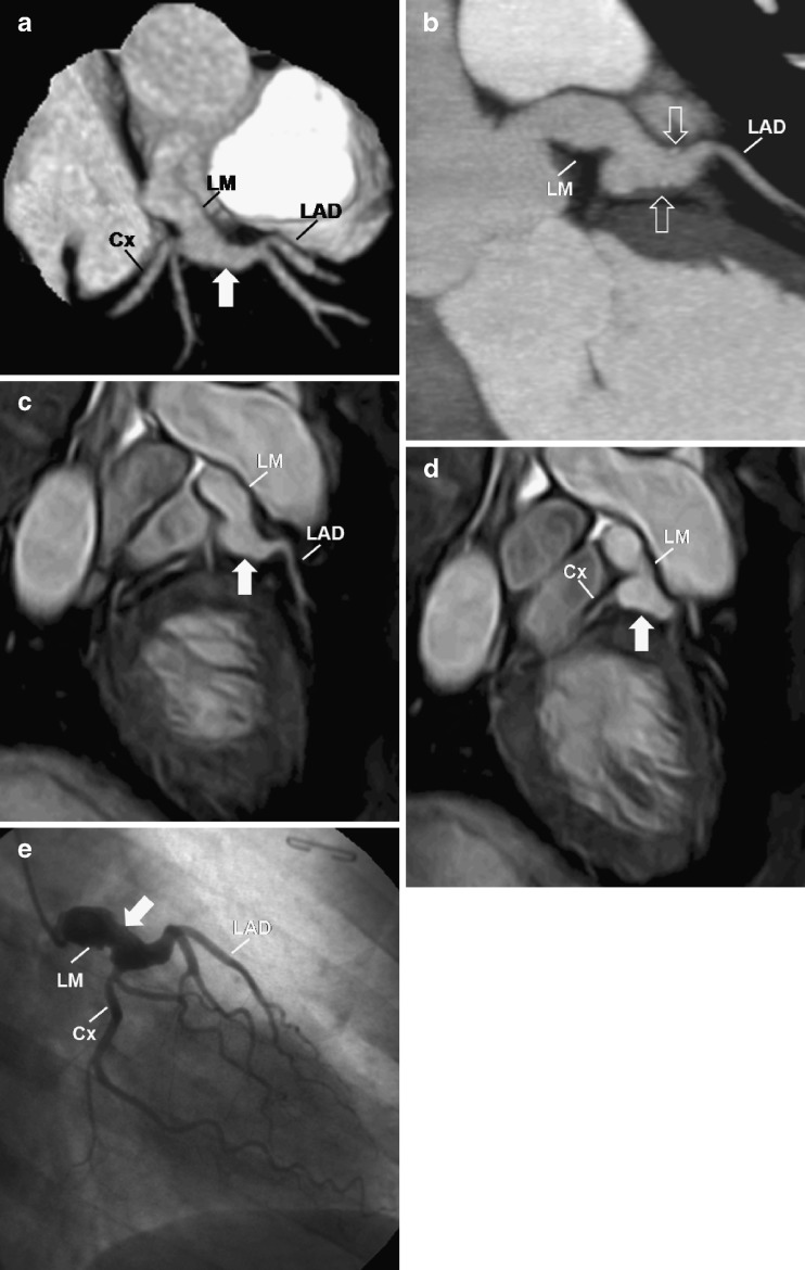
Kawasaki disease (KD) is an acute systemic vasculitis that is currently the leading cause of acquired heart disease in childhood in the United States. Cardiovascular complications are the major cause of morbidity, are responsible for virtually all deaths from KD and should be evaluated as soon as possible after the acute phase to establish the baseline status, in order to predict disease progression and determine adequate treatment. In selected patients, electrocardiography (ECG)-gated cardiac computed tomography (CT) and magnetic resonance (MR) imaging are valuable non-invasive techniques that can be used to help diagnose the cardiovascular complications associated with KD. In this article, we review the epidemiology, aetiology and pathogenesis, histopathology, clinical features, cardiovascular complications and imaging, focusing on the role of cardiac CT and MR on the initial assessment and follow-up of the cardiovascular complications of KD.


Figures

Fig. 1
Fig. 1
A 3-year-old child with KD. Photographs show erythema, fissuring and bleeding of the lips (a) and diffuse maculopappular erythematous rash predominating the trunk and neck, with evident skin desquamation (b, c, d)
Fig. 2
Fig. 2
A 5-year-old boy with KD. Reconstructed volume-rendered 3D CT coronary angiography (a, b) showing a fusiform aneurysm (arrow) involving the middle segment of the right coronary artery (RCA)
Fig. 3
Fig. 3
A 5-year-old boy with KD. Reconstructed volume-rendered 3D CT coronary angiography (a, b, c) revealing coronary arteries dilatations involving the right coronary artery (RCA), including the posterior descending artery (PD), the left main (LM) and the proximal segments of the left anterior descending artery (LAD) and circumflex artery (Cx) and the ramus intermedium (RI). Maximum-intensity projection 2D curved planar reformatting CT angiography of the RCA (d) showing diffuse ectasia (arrow) affecting all segments, including the PD. Maximum-intensity projection 2D curved planar reformatting CT angiography of the left coronary artery (e, f, g) revealing a giant coronary without intraluminal thrombus of the LM and proximal segments of the LAD, Cx and RI (arrows)
Fig. 4
Fig. 4
A 40-year-old woman with a history of KD. Reconstructed volume-rendered 3D CT coronary angiography (a) demonstrating the left coronary artery with a giant fusiform aneurysm (arrow). Two-dimensional curved planar reformatting CT angiography (b) showing a giant fusiform aneurysm (arrow) 9 mm in diameter in the left main (LM) coronary artery and minimal mural calcifications (open arrows). MR coronary angiogram (c, d) and conventional coronary angiogram (e) showing the LM giant aneurysm (arrow). LAD left anterior descending, Cx left circumflex, RCA right coronary artery
Fig. 5
Fig. 5
A 16-year-old male patient with a history of KD. Reconstructed maximum-intensity-projection 3D MR angiography (a) demonstrating bilateral fusiform aneurysms in the brachial arteries (arrows). MR coronary angiograms (b, c, d, e) with the maximum-intensity-projection technique showing normal coronaries, without evidence of aneurysms

Similar articles

Cited by

References

    1. Newburger JW, Takahashi M, Gerber MA, Gewitz MH, Tani LY, Burns JC, et al. Diagnosis, treatment, and long-term management of Kawasaki disease: a statement for health professionals from the Committee on Rheumatic Fever, Endocarditis and Kawasaki Disease, Council on Cardiovascular Disease in the Young, American Heart Association. Circulation. 2004;110:2747–2771. doi: 10.1161/01.CIR.0000145143.19711.78. - DOI - PubMed
    1. Rowley AH, Shulman ST. Kawasaki syndrome. Clin Microbiol Rev. 1998;11:405–414. - PMC - PubMed
    1. Chung C, Stein L. Kawasaki disease: a review. Radiology. 1998;208:25–33. - PubMed
    1. Burns JC. The riddle of Kawasaki disease. N Engl J Med. 2007;356:659–661. doi: 10.1056/NEJMp068268. - DOI - PubMed
    1. Burns JC, Kushner HI, Bastian JF, Shike H, Shimizu C, Matsubara T, Turner CL. Kawasaki disease: a brief history. Pediatrics. 2000;106:E27. doi: 10.1542/peds.106.2.e27. - DOI - PubMed

LinkOut - more resources