. 2011 Feb;2(1):69-84.
doi: 10.1007/s13244-010-0051-6. Epub 2010 Dec 2.

CT findings of misleading features of colonic diverticulitis

Affiliations

CT findings of misleading features of colonic diverticulitis

Ismahen Ben Yaacoub et al. Insights Imaging. 2011 Feb.

Abstract

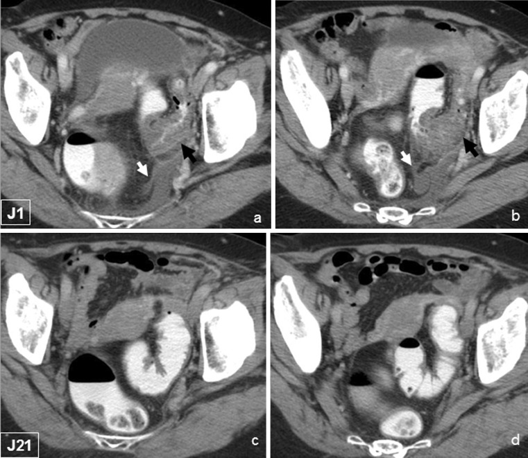
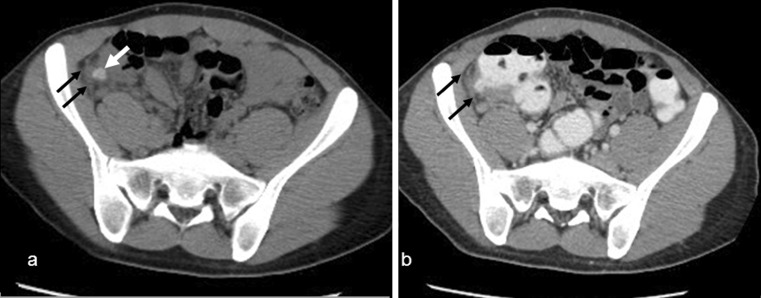
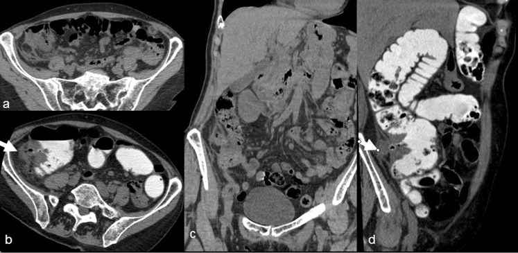
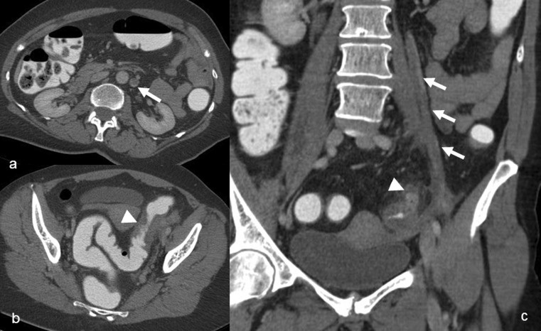
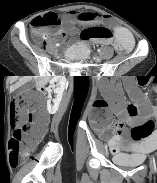
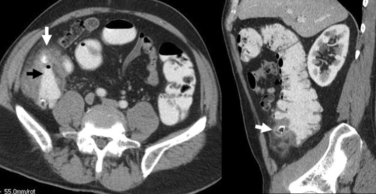
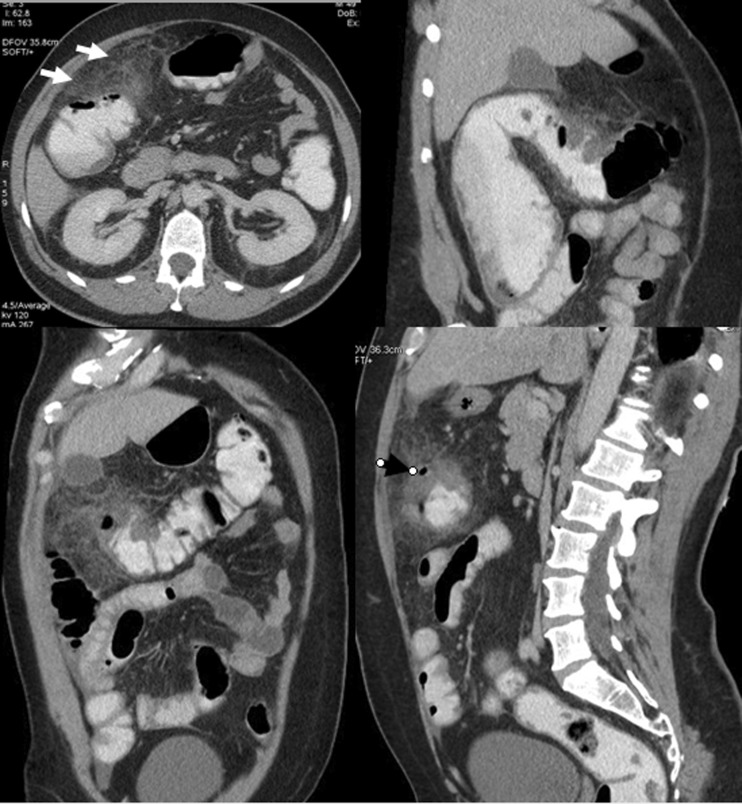
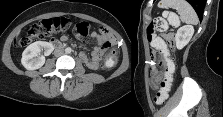
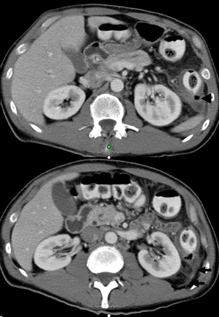
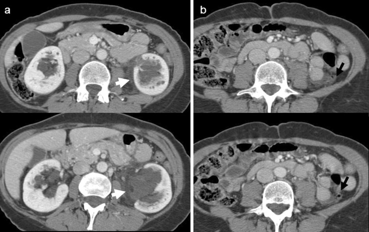
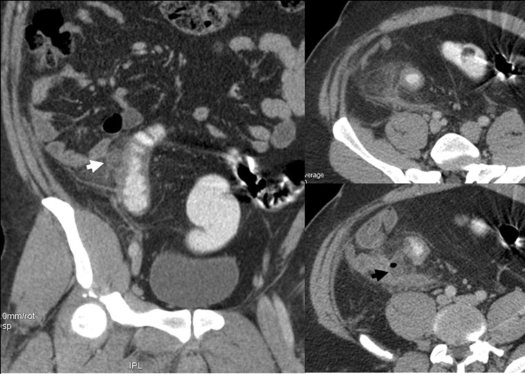
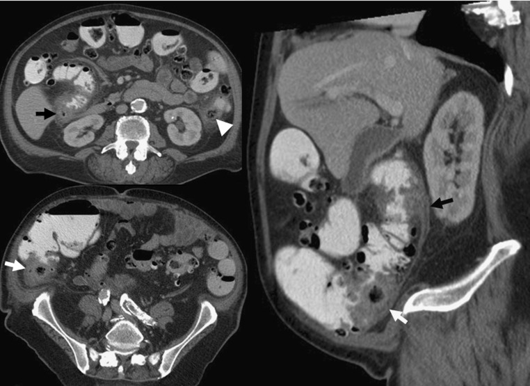
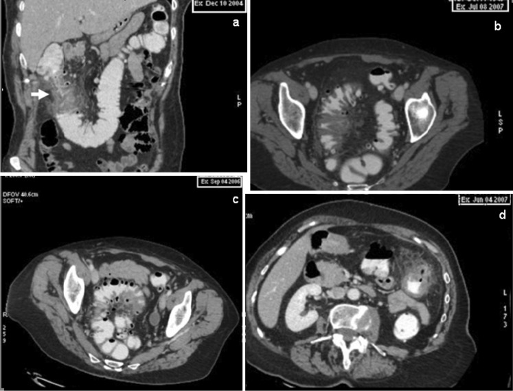
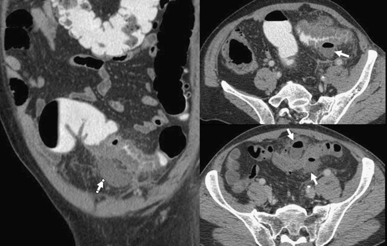
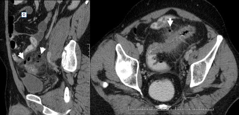
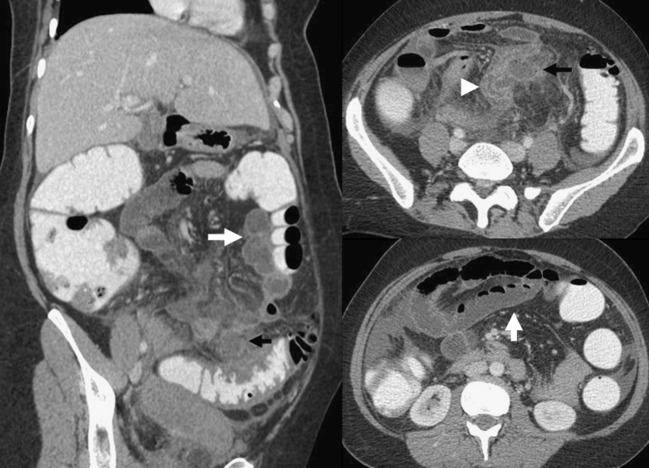
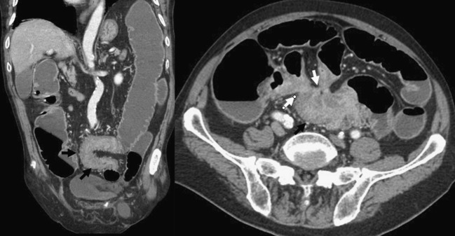
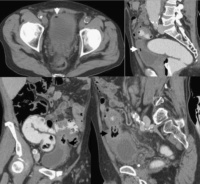
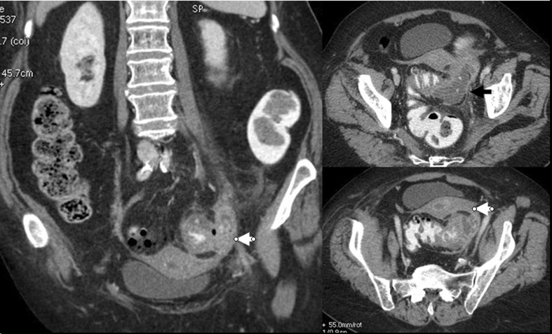
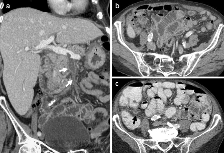
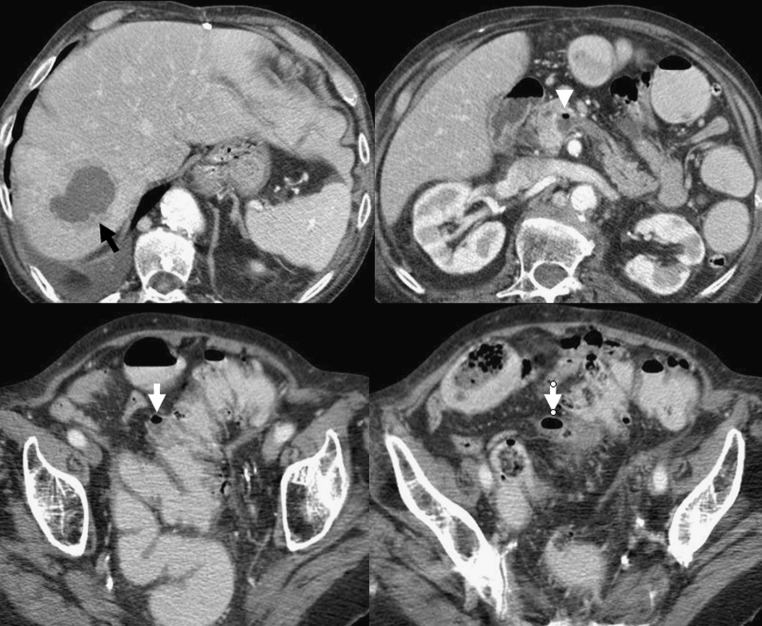
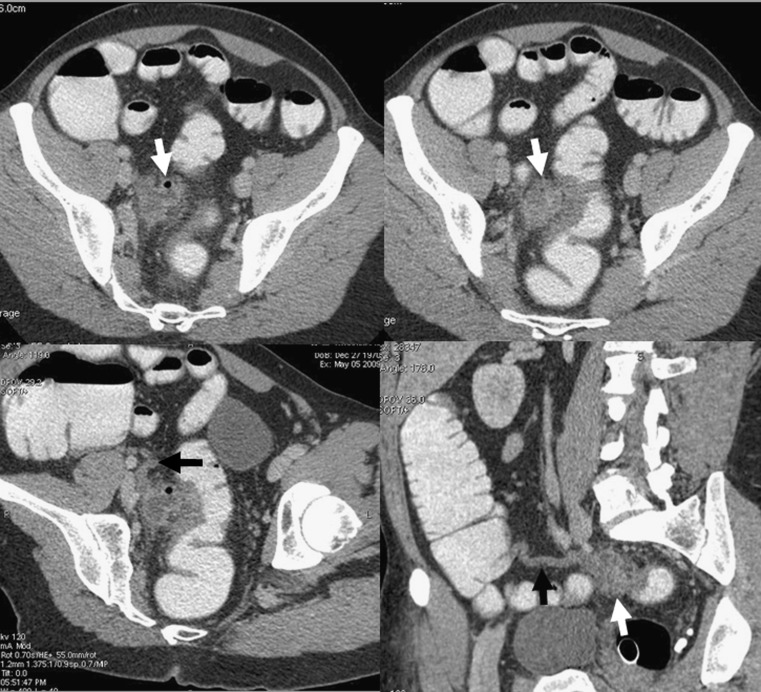
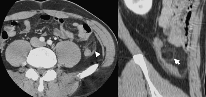
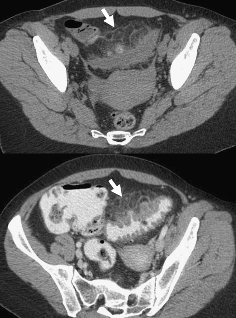
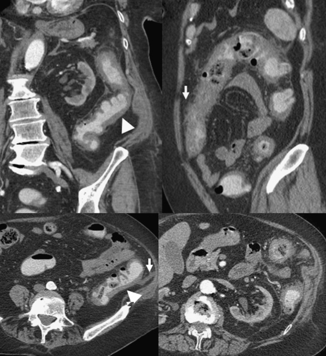
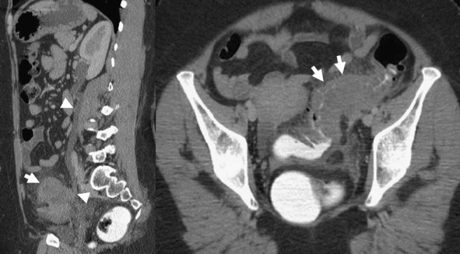
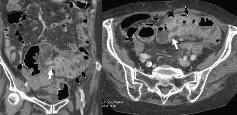
Colonic diverticulitis (CD) is a common entity whose diagnosis is particularly based on computed tomography (CT) examination, which is the imaging technique of choice. However, unusual CT findings of CD may lead to several difficulties and potential pitfalls: due to technical errors in the management of the CT examination, due to the anatomical situation of the diseased colon, in diagnosing unusual complications that may concern the gastrointestinal tract, intra- and retroperitoneal viscera or the abdominal wall, and in differentiating CD from other abdominal inflammatory and infectious conditions or colonic cancer. The aim of this work is to delineate the pitfalls of CT imaging and illustrate misleading CT features in patients with suspected CD.


Figures

Fig. 1
Fig. 1
Abdominal CT performed within 24 h of symptoms onset demonstrating typical features of sigmoid diverticulitis (a, b), with colonic wall thickening (black arrow) and paracolic inflammation (white arrow). CT performed three weeks later, does not show any suggestion of diverticulitis
Fig. 2
Fig. 2
Stercolith within a diverticulum of the right colon is more clearly identified on non-contrast-enhanced CT (a) (white arrow) than on contrast-enhanced CT (b). The association with mild paracolic inflammation (black arrows) is diagnostic for diverticulitis of the right colon
Fig. 3
Fig. 3
Diverticulitis of the right colon in a 67-year-old woman with a history of renal failure. Only non-contrast-enhanced CT was performed in this patient and the assessment of CT features of diverticulitis (white arrows) was feasible thanks to colonic opacification
Fig. 4
Fig. 4
Abdominal CT in a 63-year-old woman, demonstrating typical features of non-complicated sigmoid diverticulitis (arrowheads). However, left uterine vein thrombosis with vein enlargement and filling defect (white arrows) was detected on portal phase contrast-enhanced CT
Fig. 5
Fig. 5
Diverticulitis of the right colon, with an inflamed diverticulum of the posterior wall of the caecum with fat stranding (arrow). MPR showing adjacent normal appendix (arrowheads) confirmed the diagnosis
Fig. 6
Fig. 6
Diverticulitis of the right colon with an inflamed anterior diverticulum (white arrow) and thickening of the caecal wall, demonstrating the “arrowhead sign” (black arrow)
Fig. 7
Fig. 7
A 49-year-old patient, presented with acute pain of the upper right quadrant of the abdomen. CT showed a significant fat densification of the upper right quadrant (white arrows) mimicking segmental omental infarction. However, thickening of the right transverse colon associated with a typical “inflamed diverticulum” (black arrow) were diagnostic for diverticulitis of the transverse colon
Fig. 8
Fig. 8
Diverticulitis of the descending colon with an anterior “inflamed diverticulum” (white arrows) associated with intraperitoneal paracolic inflammation
Fig. 9
Fig. 9
Diverticulitis of the descending colon with a posterior “inflamed diverticulum” (arrowhead) associated with retroperitoneal inflammation (arrows)
Fig. 10
Fig. 10
Contrast-enhanced abdominal CT performed for acute left-sided abdominal pain in a 56-year-old woman. a Upper sections show perirenal fat stranding (white arrows). Bilateral incidental parapyelic cysts are present. b Lower sections revealed “inflamed diverticulum” within the posterior wall of the descending colon (black arrow) confirming the diagnosis of left colonic diverticulitis with almost purely retroperitoneal involvement
Fig. 11
Fig. 11
Sigmoid diverticulitis in a 29-year-old patient with a sigmoid loop lying in the right iliac fossa masquerading clinically as appendicitis. Inflamed diverticulum (white arrow). Pericolic abscess (black arrow)
Fig. 12
Fig. 12
Synchronous multifocal diverticulitis with three “inflamed diverticula” visible on the same CT examination: two of them are located in the right ascending colon (white and black arrows) and one in the left descending colon (arrowhead)
Fig. 13
Fig. 13
a A 61-year-old patient was admitted to hospital in December 2004 with acute abdominal pain. Abdominal CT was diagnostic for diverticulitis of the right transverse colon. The patient was scheduled for surgery and right colonic resection was performed two months after recovery. b The patient was readmitted in September 2006, for similar clinical symptoms. The diagnosis of sigmoid diverticulitis was made on CT. Conservative treatment was performed. c The patient was readmitted in June 2007 for similar clinical symptoms. CT revealed diverticulitis involving the descending colon. Conservative treatment was performed. d The patient was readmitted in July 2007, with a fourth episode of diverticulitis involving the sigmoid colon
Fig. 14
Fig. 14
Complicated sigmoid diverticulitis with two paracolic abscesses (white arrows)
Fig. 15
Fig. 15
Diverticular perforation complicating sigmoid diverticulitis in a 35-year-old man. CT demonstrated free gas within the mesosigmoid inflamed fat (white arrows)
Fig. 16
Fig. 16
Sigmoid diverticulitis with abscess formation (black arrows) associated with thickening of adjacent small bowel wall (white arrowhead) responsible for intestinal obstruction (white arrows)
Fig. 17
Fig. 17
Chronic inflammatory narrowing of the sigmoid colon (black arrows), due to recurrent diverticulitis and responsible for large bowel obstruction and retraction of small bowel loops around an inflammatory mass (white arrows)
Fig. 18
Fig. 18
Contrast-enhanced abdominal CT demonstrating features of sigmoid “inflamed diverticulum” (black arrows) adjacent to a thickened bladder wall associated with free air within the bladder (white arrows), establishing the diagnosis of sigmoid diverticulitis with bladder fistulisation
Fig. 19
Fig. 19
Sigmoid diverticulitis with abscess formation (black arrow) and left adnexal extension of the inflammatory process (white arrow)
Fig. 20
Fig. 20
A 68-year-old man with abdominal pain and fever. Abdominal contrast-enhanced CT demonstrated a superior mesenteric vein thrombosis (white arrows), with no regional source identified on this first CT scan (a, b). The septic source of this pylephlebitis was diverticulitis of the right colon (black arrow) detected by a second CT examination performed later with colonic opacification (c)
Fig. 21
Fig. 21
Contrast-enhanced CT demonstrating hepatic abscess (black arrow) and pylephlebitis with intravenous free air (white arrowhead) in the superior mesenteric vein. The lower CT sections revealed the septic source which was sigmoid diverticulitis with a small paracolic abscess (white arrows)
Fig. 22
Fig. 22
Paracolic abscess with thickening of the adjacent sigmoid wall was very suggestive of sigmoid diverticulitis (white arrows). Curved MPR revealed a mild thickening of the extremity of the appendix (black arrows), which was pointing to the paracolic abscess. A final diagnosis of a perforated appendicitis was confirmed at surgery
Fig. 23
Fig. 23
Primary appendagitis of the descending colon with a fatty oval-shaped nodule (white arrow) located antero-laterally to the colon associated with mild thickening of the parietal peritoneum (white arrowhead)
Fig. 24
Fig. 24
Non-contrast-enhanced and contrast-enhanced CT revealing typical features of sigmoid diverticulitis associated with secondary appendagitis (white arrows)
Fig. 25
Fig. 25
An 81-year-old man presenting with acute abdominal pain. Abdominal CT demonstrates extensive wall thickening of a diverticular left colon (arrowheads) with “disproportionate” pericolic fat standing (arrows) and no identified “inflamed diverticulum”, suggesting the diagnosis of ischaemic colitis rather than diverticulitis
Fig. 26
Fig. 26
Sigmoid diverticulitis mimicking a sigmoid cancer (white arrows) with left ureteral involvement resulting in hydronephrosis (white arrowheads)
Fig. 27
Fig. 27
Abdominal CT in an 80-year-old patient admitted for acute abdominal pain and fever, demonstrating sigmoid thickening suggesting colonic cancer (white arrows) associated with the CT finding of upstream diverticulitis (black arrows). Evidence of the association between these two pathological conditions was surgically proven

Similar articles

Cited by

References

    1. Zins M, Bruel JM, Pochet P, Regent D, Loiseau D. Question 1. What is the diagnostic value of the different tests for simple and complicated diverticulitis? What diagnostic strategy should be used? Gastroentérol Clin Biol. 2007;31:3S15–3S19. - PubMed
    1. Ambrosetti P, Jenny A, Becker C, Terrier TF, Morel P. Acute left colonic diverticulitis—compared performance of computed tomography and water-soluble contrast enema: prospective evaluation of 420 patients. Dis Colon Rectum. 2000;43:1363–1367. doi: 10.1007/BF02236631. - DOI - PubMed
    1. Laméris W, Randen A, Bipat S, Bossuyt PMM, Boermeester MA. Graded compression ultrasonography and computed tomography in acute colonic diverticulitis: meta-analysis of test accuracy. Eur Radiol. 2008;18:2498–2511. doi: 10.1007/s00330-008-1018-6. - DOI - PubMed
    1. Balthazar EJ, Megibow A, Schinella RA, Gordon R. Limitations in the CT diagnosis of acute diverticulitis: comparison of CT, contrast enema, and pathologic findings in 16 patients. AJR Am J Roentgenol. 1990;154:281–285. - PubMed
    1. Brengman ML, Otchy DP. Timing of computed tomography in acute diverticulitis. Dis Colon Rectum. 1998;41:1023–1028. doi: 10.1007/BF02237394. - DOI - PubMed

LinkOut - more resources