. 2011 Apr;2(2):149-159.
doi: 10.1007/s13244-010-0057-0. Epub 2011 Jan 19.

Dual- and multi-energy CT: approach to functional imaging

Dual- and multi-energy CT: approach to functional imaging

Juergen Fornaro et al. Insights Imaging. 2011 Apr.

Abstract

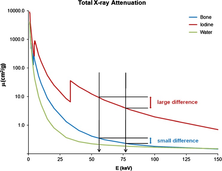
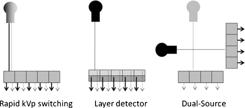
The energy spectrum of X-ray photons after passage through an absorber contains information about its elemental composition. Thus, tissue characterisation becomes feasible provided that absorption characteristics can be measured or differentiated. Dual-energy CT uses two X-ray spectra enabling material differentiation by analysing material-dependent photo-electric and Compton effects. Elemental concentrations can thereby be determined using three-material decomposition algorithms. In comparison to dual-energy CT used in clinical practice, recently developed energy-sensitive photon-counting detectors sample the material-specific attenuation curves at multiple energy levels and within narrow energy bands; the latter allows the detection of element-specific, k-edge discontinuities of the photo-electric cross section. Multi-energy CT imaging therefore is able to concurrently identify multiple materials with increased accuracy. These specific data on material distribution provide information beyond morphological CT, and approach functional imaging. This article reviews the principles of dual- and multi-energy CT imaging, hardware approaches and clinical applications.


Figures

Fig. 1
Fig. 1
Material-specific attenuation curves of bone, iodine and water plotted versus photon energy. Note the difference in attenuation between the mean energy of the spectra of 80 kVp and 140 kVp of iodine compared with bone
Fig. 2
Fig. 2
Illustration of different hardware approaches to dual-energy CT imaging. In rapid kVp switching the X-ray tube voltage is rapidly modulated to different kVp levels, producing spectra of lower and higher energies (left). Energy-sensitive layer detectors are superimposed on the other (middle). The top-layer detector absorbs lower energy X-ray photons, whereas the bottom-layer detector detects higher energy X-ray photons. The dual-source CT approach uses two X-ray tubes and corresponding detectors arranged at an angular off-set (right). Both X-ray tubes are operated at different kVp levels, allowing simultaneous dual-energy data acquisition
Fig. 3
Fig. 3
Volume rendering of dual-energy cranial CT angiography demonstrates arteriovenous malformation in the right frontal lobe (A, black arrow). Automated bone removal is precisely achieved using dual-energy (B), allowing improved delineation of the malformation together with drainage into the superior sagittal sinus
Fig. 4
Fig. 4
Dual-energy CT virtual non-calcium grey-scale (A) and colour-coded images (B) clearly demonstrate post-traumatic bone bruise as proven by T2-weighted magnetic resonance imaging (C). Note the intact osseous structures (D)
Fig. 5
Fig. 5
Non-contrast-enhanced abdominal dual-energy CT accurately depicts a urinary stone in the right distal ureter (A). Dual-energy postprocessing demonstrates the stone to be completely composed of non-uric acid as indicated by blue colour coding (B, arrow). None of the partitions of the stone consists of uric acid (red colour coding, not shown)
Fig. 6
Fig. 6
Weighted dual-energy image (A) averaged between low and high kVp images similar in appearance to conventional single-energy CT images. Dual-energy-derived material-specific information is used to quantify contrast material, allowing its subtraction to generate virtual non-enhanced images (B). Iodine maps show the contrast material content superimposed on grey scale CT (C). Note comparison of weighted dual-energy image with virtual non-enhanced image as well as iodine maps allowing detection or exclusion of enhancement from a single phase acquisition (see renal cyst, arrows)
Fig. 7
Fig. 7
Pulmonary dual-energy CT angiography allows assessment of lung perfusion in patients with pulmonary emphysema. Note the patchy, heterogeneous perfusion pattern mostly accentuated in the right upper lobe as demonstrated by transverse (A) and coronal (B) dual-energy CT reformations being in good agreement with perfusion scintigraphy (C)
Fig. 8
Fig. 8
Multi-energy CT allows differentiation of iodine, barium and bone in mouse. Single-energy image demonstrates similar attenuation values of iodine, barium and bone, which cannot be distinguished on the basis of CT numbers at energies >23 keV (A). Multi-energy CT data enable differentiation of iodine in circulation (red colour coding), barium in lungs (yellow) and bone (white) on transverse (B) and volume-rendered CT images (C). Note iodine and barium both have high Z-numbers and are solely differentiated on the basis of k-edges being 4 keV apart. Left lower bronchus (LLB), left lower lobe (LLL), pulmonary trunk (PA), right atrium (RA), right lower lobe (RLL), right ventricle (RV)

References

    1. Alvarez RE, Macovski A. Energy-selective reconstructions in X-ray computerized tomography. Phys Med Biol. 1976;21:733–744. doi: 10.1088/0031-9155/21/5/002. - DOI - PubMed
    1. Avrin DE, Macovski A, Zatz LE. Clinical application of Compton and photo-electric reconstruction in computed tomography: preliminary results. Invest Radiol. 1978;13:217–222. doi: 10.1097/00004424-197805000-00007. - DOI - PubMed
    1. Anderson NG, Butler AP, Scott NJ, Cook NJ, Butzer JS, Schleich N, Firsching M, Grasset R, Ruiter N, Campbell M, Butler PH. Spectroscopic (multi-energy) CT distinguishes iodine and barium contrast material in MICE. Eur Radiol. 2010;20:2126–2134. doi: 10.1007/s00330-010-1768-9. - DOI - PubMed
    1. Boll DT, Patil NA, Paulson EK, Merkle EM, Nelson RC, Schindera ST, Roessl E, Martens G, Proksa R, Fleiter TR, Schlomka JP. Focal cystic high-attenuation lesions: characterization in renal phantom by using photon-counting spectral CT-improved differentiation of lesion composition. Radiology. 2009;254:270–276. doi: 10.1148/radiol.09090068. - DOI - PubMed
    1. Roessl E, Proksa R. K-edge imaging in x-ray computed tomography using multi-bin photon counting detectors. Phys Med Biol. 2007;52:4679–4696. doi: 10.1088/0031-9155/52/15/020. - DOI - PubMed

LinkOut - more resources