. 2011 Jun;2(3):235-246.
doi: 10.1007/s13244-011-0085-4. Epub 2011 Mar 25.

The future of hybrid imaging-part 3: PET/MR, small-animal imaging and beyond

The future of hybrid imaging-part 3: PET/MR, small-animal imaging and beyond

Thomas Beyer et al. Insights Imaging. 2011 Jun.

Erratum in

Abstract

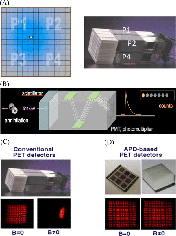
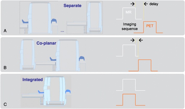
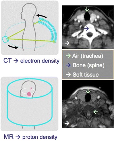
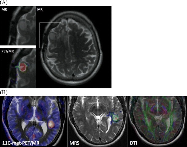
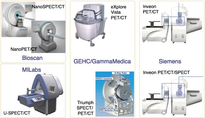
Since the 1990s, hybrid imaging by means of software and hardware image fusion alike allows the intrinsic combination of functional and anatomical image information. This review summarises in three parts the state of the art of dual-technique imaging with a focus on clinical applications. We will attempt to highlight selected areas of potential improvement of combined imaging technologies and new applications. In this third part, we discuss briefly the origins of combined positron emission tomography (PET)/magnetic resonance imaging (MRI). Unlike PET/computed tomography (CT), PET/MRI started out from developments in small-animal imaging technology, and, therefore, we add a section on advances in dual- and multi-modality imaging technology for small animals. Finally, we highlight a number of important aspects beyond technology that should be addressed for a sustained future of hybrid imaging. In short, we predict that, within 10 years, we may see all existing multi-modality imaging systems in clinical routine, including PET/MRI. Despite the current lack of clinical evidence, integrated PET/MRI may become particularly important and clinically useful in improved therapy planning for neurodegenerative diseases and subsequent response assessment, as well as in complementary loco-regional oncology imaging. Although desirable, other combinations of imaging systems, such as single-photon emission computed tomography (SPECT)/MRI may be anticipated, but will first need to go through the process of viable clinical prototyping. In the interim, a combination of PET and ultrasound may become available. As exciting as these new possible triple-technique-imaging systems sound, we need to be aware that they have to be technologically feasible, applicable in clinical routine and cost-effective.


Figures

Fig. 1
Fig. 1
a Example of photomultiplier tubes (PMT)-bismuth germanate (BGO) block detector from a clinical PET system. Readout is performed using the PMTs that are connected to the pixellated scintillator block. Light sharing is used to distribute light originating from a single pixel between the readout PMTs (P1-P4). The position of the incident annihilation photon event can be calculated using an Anger-weighting of the measured signals (b). b Schematics of the detection process from annihilation to stopping the annihilation of photons in the crystal and signal transformation inside the PMT. c Conventional PET detectors (see a) work only outside magnetic fields (B = 0). If a PMT is operated inside a magnetic field (B > 0), then the multiplier step is distorted and the readout map severely distorted. d Avalanche photodiode (APD)-based detectors are semiconductors that can be operated in magnetic fields, even at higher field strengths. Images courtesy Prof. B. Pichler, Tübingen
Fig. 2
Fig. 2
Different designs for combined clinical PET/MR systems: (A) patients can be shuttled between separate MR and PET(/CT) systems operated in different rooms, (B) patients are positioned on a common table platform between stationary PET and MR systems; the delay between the MR- and PET-examination is reduced (Philips Healthcare), and (C) patients are positioned inside an integrated PET/MR gantry (Siemens Healthcare) with a PET insert that is mounted within a whole-body MR offering simultaneous PET/MR acquisitions
Fig. 3
Fig. 3
MR-based attenuation correction is demanding as the appearance of air (turquoise arrow) and bone (blue arrow) on MR images is very similar despite their significantly different attenuation coefficients for ionising radiation (see CT, top)
Fig. 4
Fig. 4
a Patient with meningioma in the right frontal lobe. Axial MR and simultaneous PET/MR images through the lesion: T2-weighted MRI, 68Ga-DOTATOC PET. b A 42-year-old man with a neurocytoma. PET/MR images were acquired simultaneously following injections of 11C-methionine (left). Simultaneously acquired chemical shift imaging MRS provides a map of the choline to N-acetyl-aspartate ratio (centre). Simultaneous diffusion tensor imaging (DTI) shows the clear relationship with the adjacent optic radiation. Cases courtesy of Drs. Boss, Bisdas and Schwenzer (UH Tübingen, Department of Radiology)
Fig. 5
Fig. 5
Different design concepts for dual- and triple-technique imaging systems for pre-clinical applications. In general, system designs are similar to clinical dual-technique imaging systems even for the docking triple-technique system shown in the right panel
Fig. 6
Fig. 6
Alternative combinations of imaging techniques in prototype designs and research testing: (a) scintimmamography imaging [58], (b) combined X-ray/ultrasound imaging [59], (c) mammotomography [60] and (d) SPECT/MRI [42]

References

    1. Padhani A, Miles K. Multiparametric imaging of tumor response to therapy. Radiology. 2010;256(2):348–364. doi: 10.1148/radiol.10091760. - DOI - PubMed
    1. Hammer B, Christensen N, Heil BG. Use of a magnetic field to increase the spatial resolution of positron emission tomography. Med Phys. 1994;21:1917–1920. doi: 10.1118/1.597178. - DOI - PubMed
    1. Christensen N, et al. Positron emission tomography within a magnetic field using photomultiplier tubes and lightguides. Phys Med Biol. 1995;40:691–697. doi: 10.1088/0031-9155/40/4/014. - DOI - PubMed
    1. Shao Y, et al. Development of a PET detector system compatible with MRI/NMR systems. IEEE Trans Nucl Sci. 1997;44(3):1167–1171. doi: 10.1109/23.596982. - DOI
    1. Slates R, et al. Design of a small animal MR compatible PET scanner. IEEE Trans Nucl Sci. 1999;46:565–570. doi: 10.1109/23.775580. - DOI