. 2011 Jun;2(3):281-295.
doi: 10.1007/s13244-011-0072-9. Epub 2011 Feb 11.

CT imaging of blunt chest trauma

Affiliations

CT imaging of blunt chest trauma

Anastasia Oikonomou et al. Insights Imaging. 2011 Jun.

Abstract

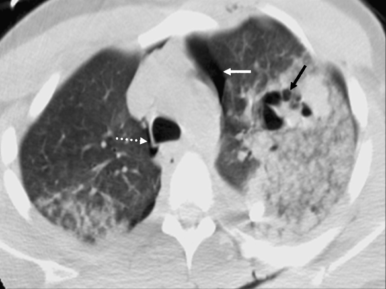
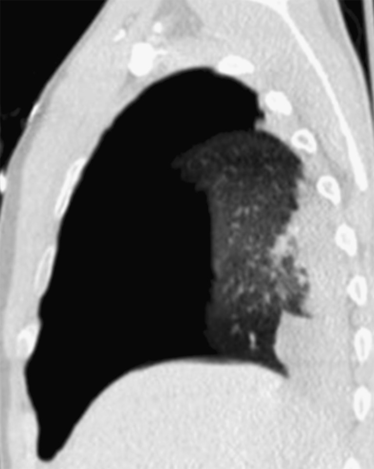
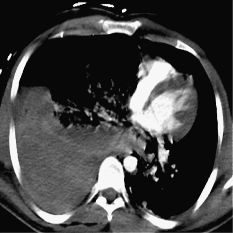
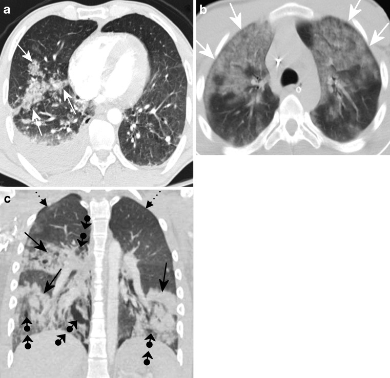
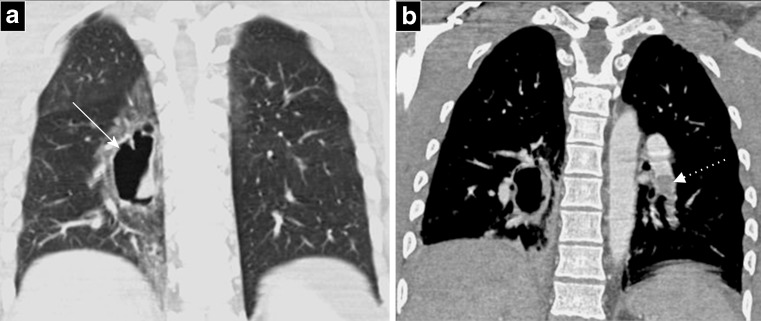
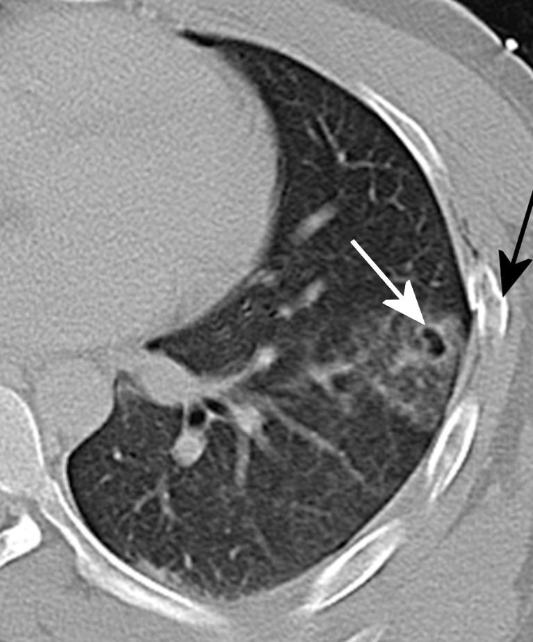
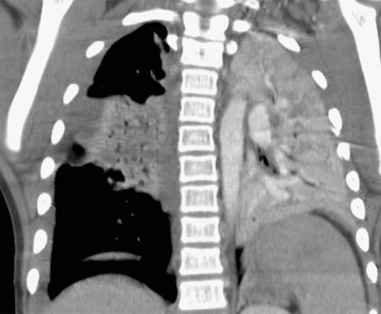
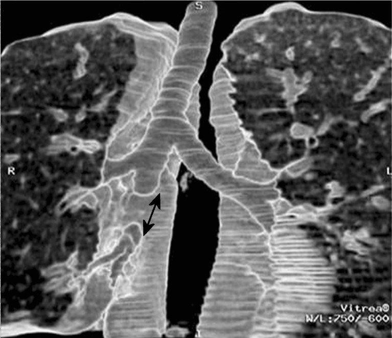
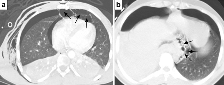
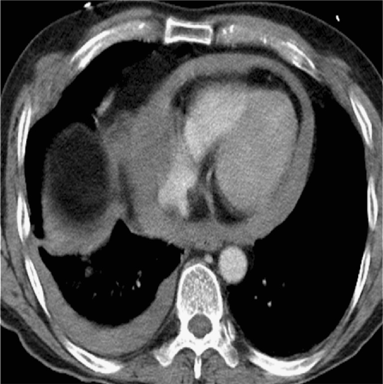
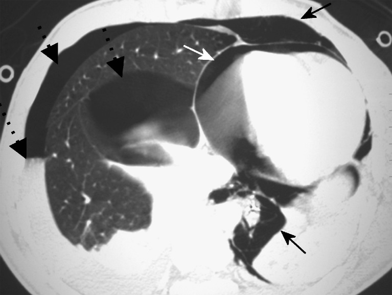
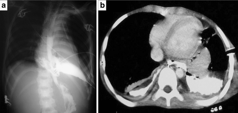
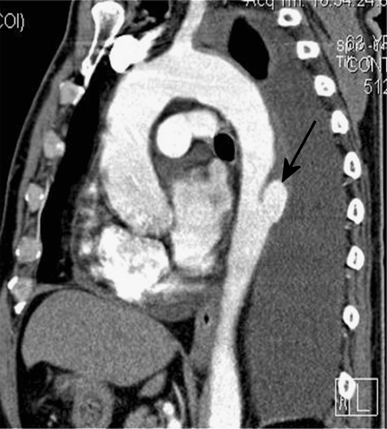
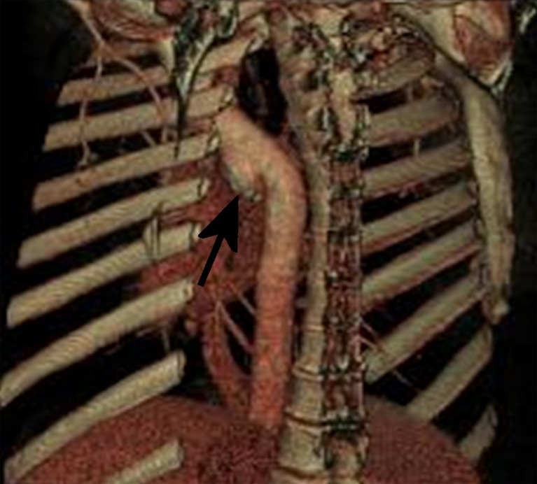
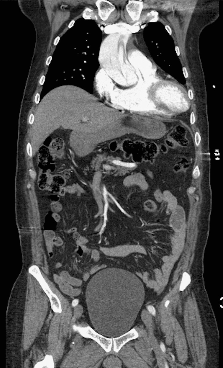
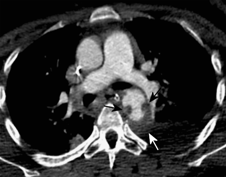
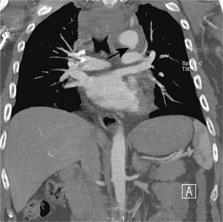
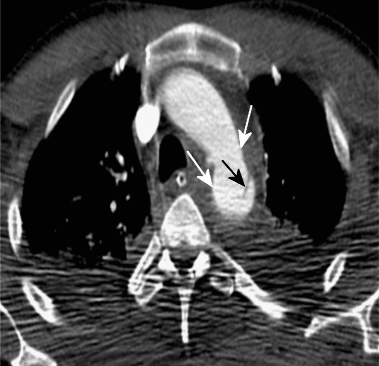
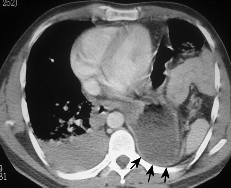
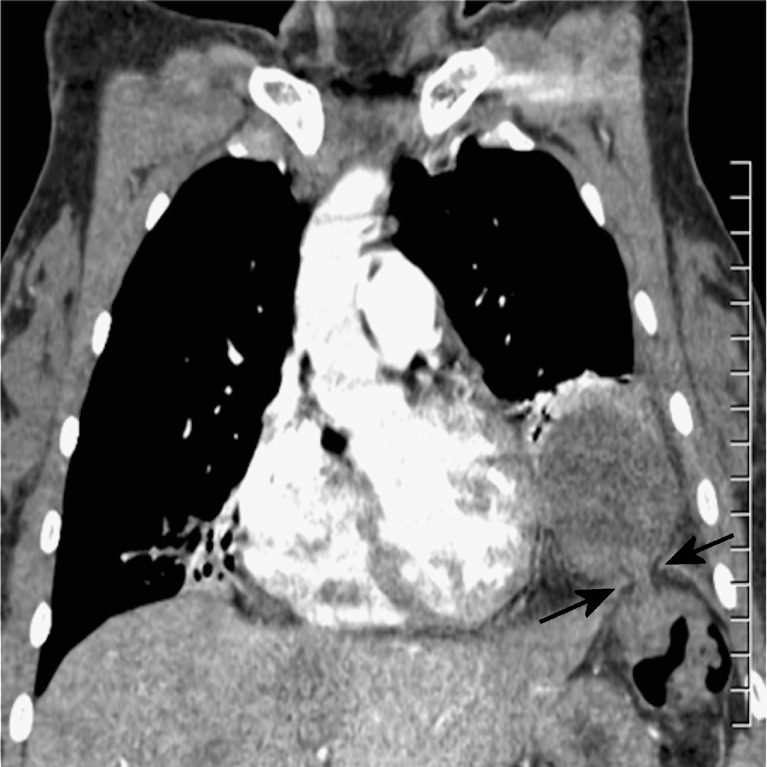
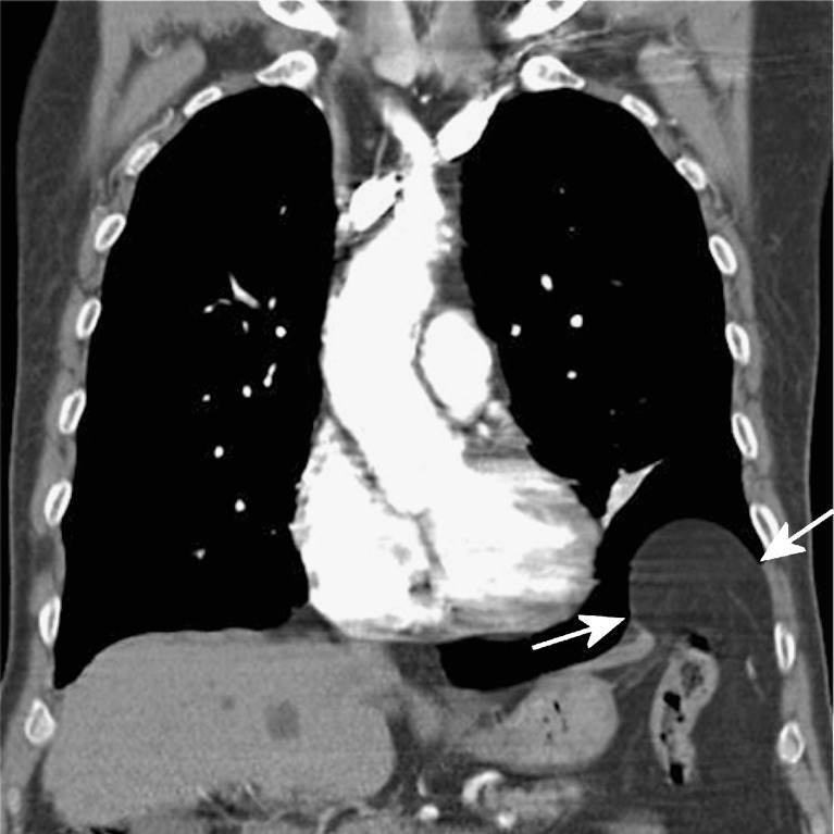
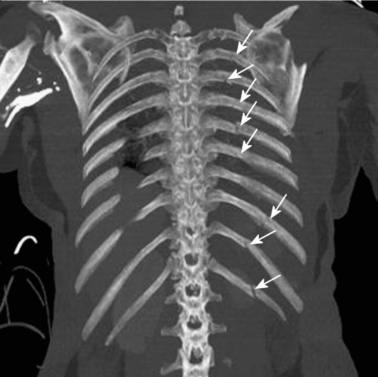
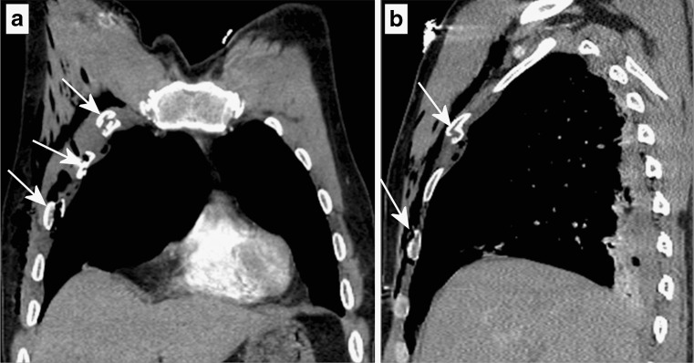
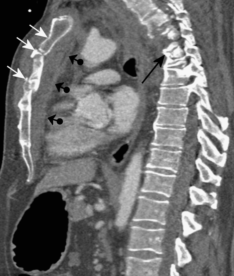
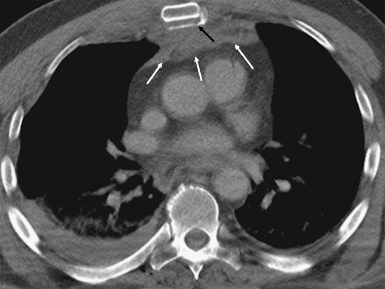
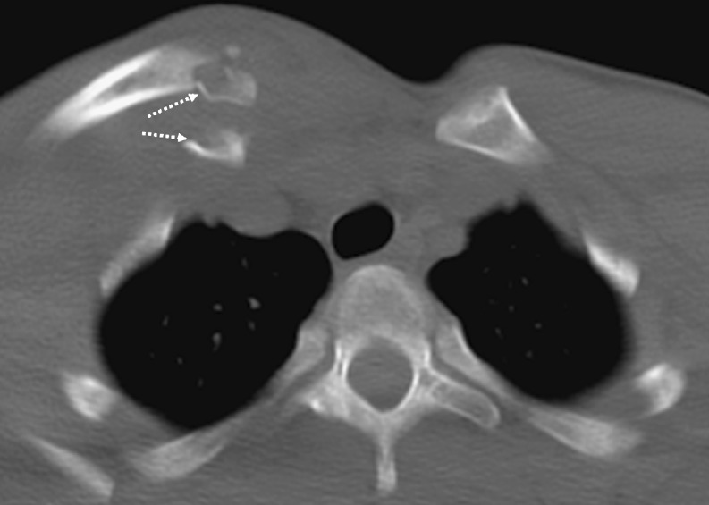
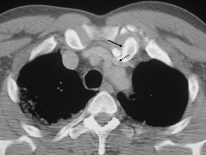
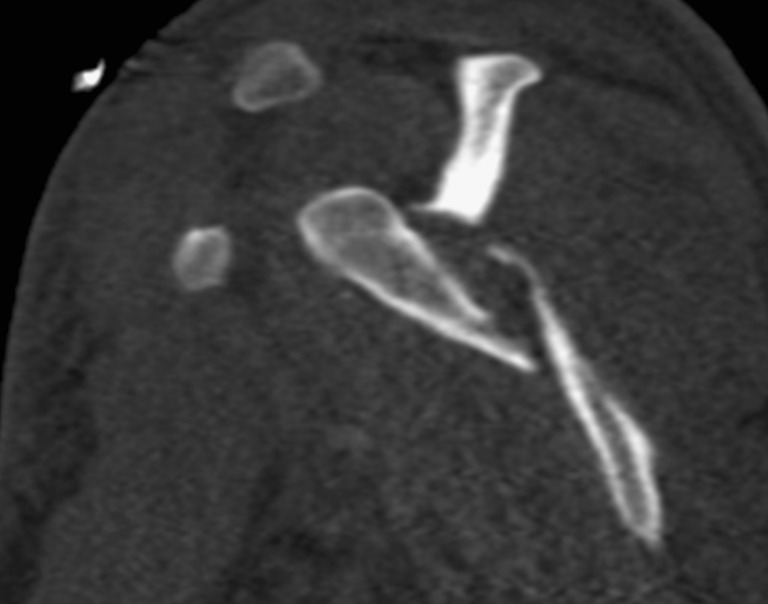
BACKGROUND: Thoracic injury overall is the third most common cause of trauma following injury to the head and extremities. Thoracic trauma has a high morbidity and mortality, accounting for approximately 25% of trauma-related deaths, second only to head trauma. More than 70% of cases of blunt thoracic trauma are due to motor vehicle collisions, with the remainder caused by falls or blows from blunt objects. METHODS: The mechanisms of injury, spectrum of abnormalities and radiological findings encountered in blunt thoracic trauma are categorised into injuries of the pleural space (pneumothorax, hemothorax), the lungs (pulmonary contusion, laceration and herniation), the airways (tracheobronchial lacerations, Macklin effect), the oesophagus, the heart, the aorta, the diaphragm and the chest wall (rib, scapular, sternal fractures and sternoclavicular dislocations). The possible coexistence of multiple types of injury in a single patient is stressed, and therefore systematic exclusion after thorough investigation of all types of injury is warranted. RESULTS: The superiority of CT over chest radiography in diagnosing chest trauma is well documented. Moreover, with the advent of MDCT the imaging time for trauma patients has been significantly reduced to several seconds, allowing more time for appropriate post-diagnosis care. CONCLUSION: High-quality multiplanar and volumetric reformatted CT images greatly improve the detection of injuries and enhance the understanding of mechanisms of trauma-related abnormalities.


Figures

Fig. 1
Fig. 1
Occult pneumothorax. Axial CT at lung window shows a small occult pneumothorax (missed on chest radiography) at the anteromedial part of the left upper lobe (white arrow). There are also bilateral ground-glass opacities and areas of consolidation in both upper lobes consistent with lung contusions. Note the presence of numerous small confluent pneumatoceles (Swiss cheese appearance) in the anterior segment of the left upper lobe, consistent with lung laceration (black arrow). Note also the presence of small pneumomediastinum (dotted white arrow)
Fig. 2
Fig. 2
Tension pneumothorax. Sagittal reformatted CT image at lung window showing tension pneumothorax with significantly collapsed lung at the posterior part of the hemithorax associated with ipsilateral pleural effusion
Fig. 3
Fig. 3
Tension haemopneumothorax. Axial contrast-enhanced CT at mediastinal window shows a right tension haemopneumothorax with heterogeneous increased density due to presence of blood clots and a significant shift of the mediastinum contralaterally
Fig. 4
Fig. 4
Lung contusion. Axial (a, b) and coronal (c) CT images at lung window show nodular opacities of ground-glass opacity that do not respect the lung boundaries of the right upper lobe (white arrows) (a), diffuse areas of ground-glass opacity in the upper lobes bilaterally with subpleural sparing (white arrows) (b) and multiple areas of consolidation with air bronchograms (white arrows) and small lacerations (black arrows) in both lungs consistent with lung contusions. Note small bilateral pneumothorax in both lung apices (black dotted arrows) and cardiophrenic angles (black dotted arrows) (c)
Fig. 5
Fig. 5
Lung laceration, type II. Coronal reformatted CT image at lung window (a) shows a lobulated paraspinal pneumatocele (arrow) surrounded by ground-glass opacity (contusion) in the right lung consistent with lung laceration (type II?). On mediastinal window lung laceration is seen to have been complicated by acute pulmonary embolism (dotted arrow)
Fig. 6
Fig. 6
Lung laceration, type IV. Axial CT image of the left lung at lung window shows a small peripheral laceration (white arrow) beneath a rib fracture (black arrow) surrounded by ground-glass opacity (lung contusion) and associated with a small ipsilateral pneumothorax
Fig. 7
Fig. 7
Blast lung injury. Twenty-two-year-old patient who experienced the explosion of a grenade in his hands. Coronal CT reformatted contrast-enhanced CT image at mediastinal window shows bilateral perilar consolidations mimicking a butterfly or bat-wing appearance, consistent with blast lung. The left lung is almost completely collapsed, and there are bilateral haemothoraces
Fig. 8
Fig. 8
Bronchial transection. A 22-year-old man involved in a car accident. Volume-rendered image of the tracheobronchial tree showing complete transection of the right intermediate bronchus (two-way arrow). (Courtesy of Dr Montserrat Bret, University Hospital La Paz, Madrid)
Fig. 9
Fig. 9
Pneumomediastinum. Axial CT images at wide lung window show pneumomediastinum with the presence of septae within the air in the anterior mediastinum (black arrows) (a), and in the middle and posterior mediastinum (black arrows) (b). Note also a right pneumothorax, bilateral lower lobe atelectases and subcutaneous emphysema (a) and a right haemopneumothorax and left pneumothorax (b)
Fig. 10
Fig. 10
Haemopericardium. Axial contrast-enhanced CT of the lower thorax at mediastinal window shows haemopericardium that may represent an indirect sign of pericardial or heart injury in a polytraumatised patient after a motor vehicle accident. There is also a small right haemothorax
Fig. 11
Fig. 11
Pneumopericardium. Axial CT image at lung window shows extensive pneumopericardium (white arrow), pneumomediastinum (black arrows), haemopneumothorax (black dotted arrows), collapsed left lung with ipsilateral shift of the mediastinum and collapse of the right lower lobe
Fig. 12
Fig. 12
Traumatic oesophageal rupture. A 12-year-old boy traumatised during a fall from a tree. Oesophagogram with per os administration of water-soluble contrast medium (a) shows leakage of contrast medium into both pleural spaces. Axial CT image of the thorax at the level of the lung bases (b) verifies the leakage of the contrast medium into the left and right pleural spaces. (Image reproduced from: Arora A, Puri SK, Upreti L, et al (2010). Oesophageal rupture: a rare complication of blunt trauma, {Online}. URL: http://www.eurorad.org/case.php?id=8447)
Fig. 13
Fig. 13
Traumatic aortic pseudoaneurysm. Sagittal reformatted contrast-enhanced CT image of the thoracic aorta reveals a pseudoaneurysm of the greater curve of the mid-descending thoracic aorta (black arrow)
Fig. 14
Fig. 14
Traumatic aortic pseudoaneurysm. Three-dimensional reconstructed CT image of the thoracic aorta shows a pseudoaneurysm of the inferior curve of the thoracic aorta immediately distal to the isthmus (arrow)
Fig. 15
Fig. 15
Traumatic aortic dissection. Coronal contrast-enhanced CT reformatted image of the thorax and abdomen shows a traumatic dissection of the ascending thoracic aorta (Stanford type A)
Fig. 16
Fig. 16
Axial contrast-enhanced CT image shows thrombus protruding into the lumen of the descending thoracic aorta (black arrows), indicating aortic injury surrounded by periaortic haematoma (white arrow)
Fig. 17
Fig. 17
Minimal aortic injury. Coronal MIP CT image of the thorax shows minimal aortic injury at the aortic isthmus (black arrow) involving only the intima, surrounded by periaortic haematoma
Fig. 18
Fig. 18
Axial contrast-enhanced CT image shows presence of an intimal flap at the aortic arch (black arrow) surrounded by mediastinal haematoma with no preserved fat plane with the aorta (white arrows). Bilateral pleural haemothoraces are also seen
Fig. 19
Fig. 19
Dependent viscera sign. Axial contrast-enhanced CT at the level of the lower lobes, at mediastinal window, shows intrathoracic presence of the stomach abutting the left posterior thoracic wall without intervening in the left hemidiaphragm (black arrows)
Fig. 20
Fig. 20
The “hourglass” or “collar” sign of diaphragmatic rupture. Reformatted coronal contrast-enhanced CT image of the thorax shows waist-like stricture of the herniated left colon intrathoracically through the small defect of the left hemidiaphragm (black arrows)
Fig. 21
Fig. 21
Reformatted coronal contrast-enhanced CT image of the thorax shows intrathoracic herniation of intraperitoneal fat through the large defect of the left hemidiaphragm (white arrows)
Fig. 22
Fig. 22
Coronal MIP CT image showing multiple contiguous left rib fractures (arrows)
Fig. 23
Fig. 23
Coronal (a) and sagittal (b) reconstructed CT images show fractures of three contiguous right ribs (arrows) that were associated with paradox motion of the chest during respiration. Flail chest was suspected clinically and verified on imaging
Fig. 24
Fig. 24
Sternal fracture. Sagittal reconstructed CT image shows multiple fractures of the manubrium and the body of the sternum (white arrows) accompanied by extensive retrosternal haematoma (black ball arrows). Note also fracture of a thoracic vertebra (black arrow)
Fig. 25
Fig. 25
Sternal fracture. Axial CT image at mediastinal window shows sternal fracture associated with retrosternal haematoma (black arrow). Note the preserved fat plane with the aorta, excluding the presence of aortic injury (white arrows)
Fig. 26
Fig. 26
Anterior sternoclavicular dislocation. Axial CT image shows clavicular fracture and anterior sternoclavicular dislocation (dotted arrows)
Fig. 27
Fig. 27
Posterior sternoclavicular dislocation. Axial CT image shows posterior sternoclavicular dislocation (black arrow) associated with compression of the left innominate vein (black dotted arrow)
Fig. 28
Fig. 28
Scapular fracture. Sagittal reconstructed CT image shows multiple fractures of the left scapula (arrows)
Fig. 29
Fig. 29
Thoracic spine fracture. Coronal (a) and sagittal (b) CT reconstructed images of two different patients show fractures of the upper thoracic vertebrae with great detail
Fig. 30
Fig. 30
Thoracic spine fracture and compressive myelopathy. Sagittal T2-weighted MRI of the cervicothoracic spine undertaken 1 week after a motor vehicle accident verifies the presence of extensive compressive myelopathy (between the two white arrows with black outline) due to fractures of the second and the third thoracic vertebrae (white arrows)

References

    1. Shanmuganathan K, Matsumoto J. Imaging of penetrating chest trauma. Radiol Clin North Am. 2006;44:225–238. doi: 10.1016/j.rcl.2005.10.002. - DOI - PubMed
    1. Kaewlai R, Avery LL, Asrani AV, Novelline RA. Multidetector CT of blunt thoracic trauma. Radiographics. 2008;28:1555–1570. doi: 10.1148/rg.286085510. - DOI - PubMed
    1. Scaglione M, Pinto A, Pedrosa I, Sparano A, Romano L. Multi-detector row computed tomography and blunt chest trauma. Eur J Radiol. 2008;65:377–388. doi: 10.1016/j.ejrad.2007.09.023. - DOI - PubMed
    1. Clark DE, Fantus RJ, editors. National Trauma Data Bank (NTDB) Annual Report 2007. Chicago, IL: American College of Surgeons; 2007. pp. 1–64.
    1. Mayberry JC. Imaging in thoracic trauma: the trauma surgeon's perspective. J Thorac Imaging. 2000;15:76–86. doi: 10.1097/00005382-200004000-00002. - DOI - PubMed

LinkOut - more resources