

Radiology in paediatric non-traumatic thoracic emergencies
- PMID: 22347978
- PMCID: PMC3259402
- DOI: 10.1007/s13244-011-0113-4
Radiology in paediatric non-traumatic thoracic emergencies
Abstract
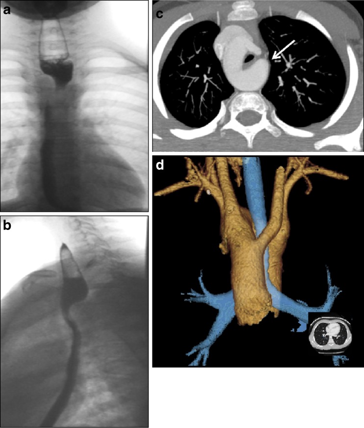
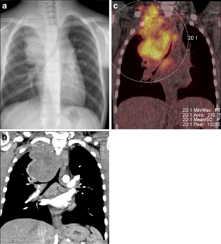
Non-traumatic thoracic emergencies in children are very frequent, and they usually present with breathing difficulties. Associated symptoms may be feeding or swallowing problems or less specific general symptoms such as fever, sepsis or chest pain. The emergencies always require a rapid diagnosis to establish a medical or surgical intervention plan, and radiological imaging often plays a key role. Correct interpretation of the radiological findings is of great importance in diagnosing and monitoring the illness and in avoiding serious complications. Plain radiography with fluoroscopy still remains the most important and frequently used tool to gain information on acute pulmonary problems. Ultrasound is the first choice for the detection and treatment of simple and complicated pleural effusions. Cross-sectional techniques such as multidetector computed tomography (MDCT) and magnetic resonance imaging (MRI) are mainly used to study pulmonary/mediastinal masses and congenital abnormalities of the great vessels and the lungs. This article will discuss the choice of imaging technique, the urgency of radiological management and the imaging characteristics of acquired and congenital causes of non-traumatic thoracic emergencies. They represent common conditions involving the respiratory tract, chest wall and the oesophagus, as well as the less frequent causes such as tumours and manifestations of congenital malformations.
Figures
Similar articles
-
[Emergency pediatric thoracic radiology].J Radiol. 2005 Feb;86(2 Pt 2):198-206. doi: 10.1016/s0221-0363(05)81347-2. J Radiol. 2005. PMID: 15798632 Review. French.
-
Chest imaging in paediatric pulmonary TB.Paediatr Respir Rev. 2020 Nov;36:65-72. doi: 10.1016/j.prrv.2020.10.002. Epub 2020 Oct 13. Paediatr Respir Rev. 2020. PMID: 33160839 Review.
-
Urgent and emergent pediatric cardiovascular imaging.Pediatr Radiol. 2025 Apr;55(4):604-621. doi: 10.1007/s00247-024-05980-y. Epub 2024 Jul 5. Pediatr Radiol. 2025. PMID: 38967787 Free PMC article. Review.
-
Imaging of Combat-Related Thoracic Trauma - Blunt Trauma and Blast Lung Injury.Mil Med. 2018 Mar 1;183(3-4):e89-e96. doi: 10.1093/milmed/usx033. Mil Med. 2018. PMID: 29514343
-
[A contribution of multidetector computed tomography to indications for chest wall stabilisation in multiple rib fractures].Acta Chir Orthop Traumatol Cech. 2011;78(3):258-61. Acta Chir Orthop Traumatol Cech. 2011. PMID: 21729644 Czech.
Cited by
-
[Radiological diagnostics of pediatric lungs].Radiologe. 2015 Jul;55(7):554-60. doi: 10.1007/s00117-014-2775-7. Radiologe. 2015. PMID: 26152499 German.
-
Acute Paediatric Tracheal Deviation and Neck Lump Secondary to Food Bolus Impaction.Cureus. 2022 Jan 24;14(1):e21553. doi: 10.7759/cureus.21553. eCollection 2022 Jan. Cureus. 2022. PMID: 35223323 Free PMC article.
-
Emergency imaging in paediatric oncology: a pictorial review.Insights Imaging. 2019 Dec 18;10(1):120. doi: 10.1186/s13244-019-0796-5. Insights Imaging. 2019. PMID: 31853747 Free PMC article. Review.
-
A leak too far--gastro-pleural fistula mimicking recurrence of repaired congenital diaphragmatic hernia following fundoplication.J Radiol Case Rep. 2013 Sep 1;7(9):33-8. doi: 10.3941/jrcr.v7i9.1505. eCollection 2013 Sep. J Radiol Case Rep. 2013. PMID: 24421956 Free PMC article.
-
Pediatric chest radiograph interpretation in a real-life setting.Eur J Pediatr. 2024 Oct;183(10):4435-4444. doi: 10.1007/s00431-024-05717-x. Epub 2024 Aug 12. Eur J Pediatr. 2024. PMID: 39133303 Free PMC article.
References
-
- Wildin SR, Chonmaitree T, Swischuk LE. Roentgenographic features of common pediatric viral respiratory tract infections. Am J Dis Child. 1988;142:43–46. - PubMed
-
- Breysem L, Loyen S, Boets A, Proesmans M, De BK, Smet MH. Pediatric emergencies: thoracic emergencies. Eur Radiol. 2002;12:2849–2865. - PubMed
-
- Frush DP, Frush KS. The ALARA concept in pediatric imaging: building bridges between radiology and emergency medicine: consensus conference on imaging safety and quality for children in the emergency setting, Feb. 23–24, 2008, Orlando, FL—executive summary. Pediatr Radiol. 2008;38(Suppl 4):S629–S632. doi: 10.1007/s00247-008-1006-7. - DOI - PubMed
LinkOut - more resources
Full Text Sources