. 2011 Dec;2(6):653-670.
doi: 10.1007/s13244-011-0122-3. Epub 2011 Aug 7.

Imaging of breast implants-a pictorial review

Imaging of breast implants-a pictorial review

Sergi Juanpere et al. Insights Imaging. 2011 Dec.

Abstract

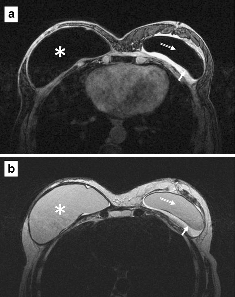
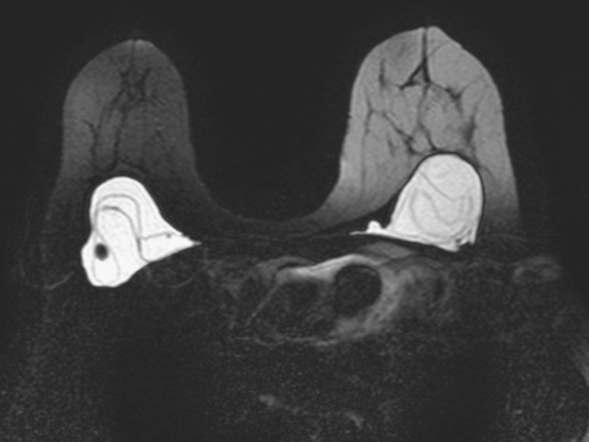
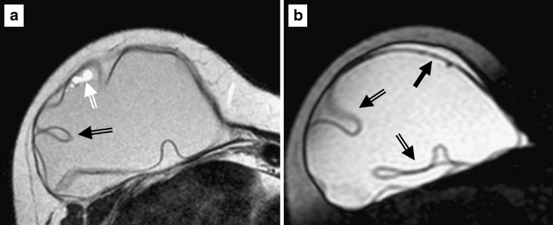
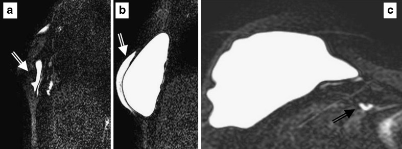
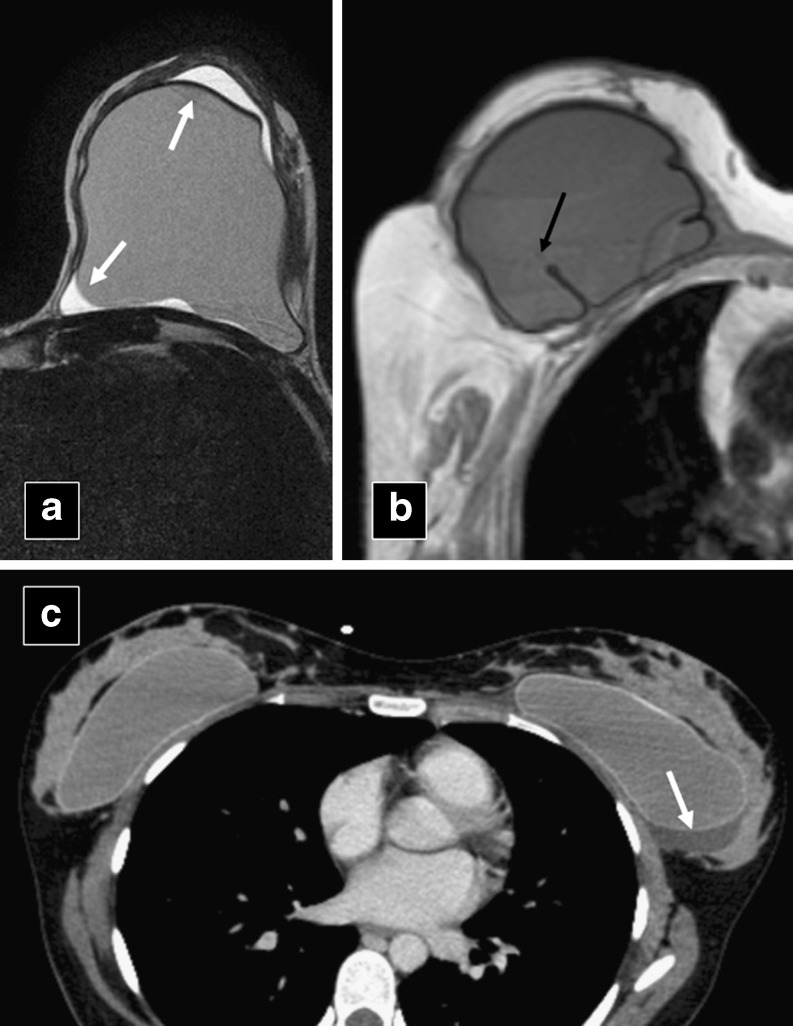
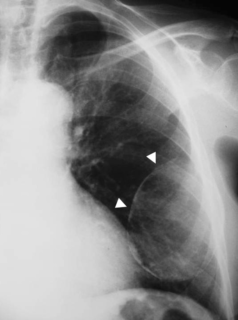
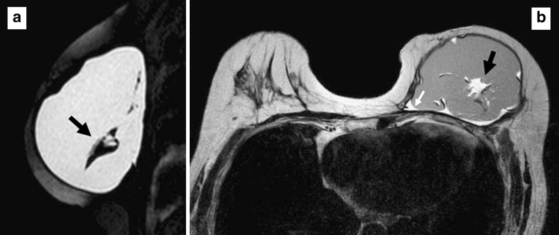
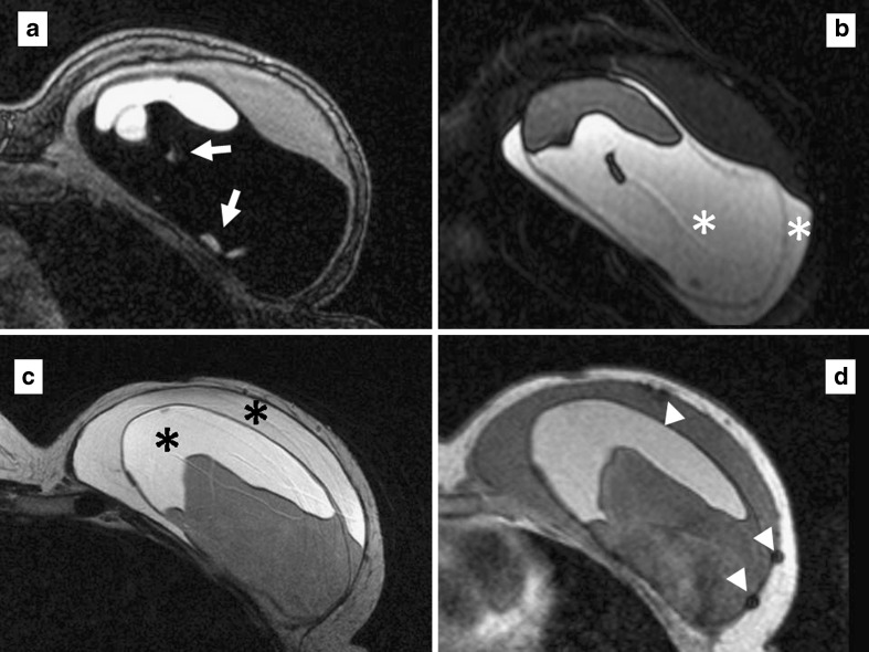
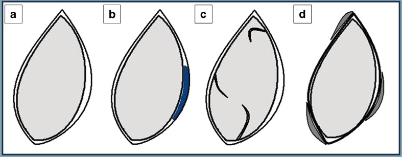
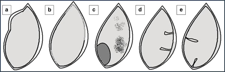
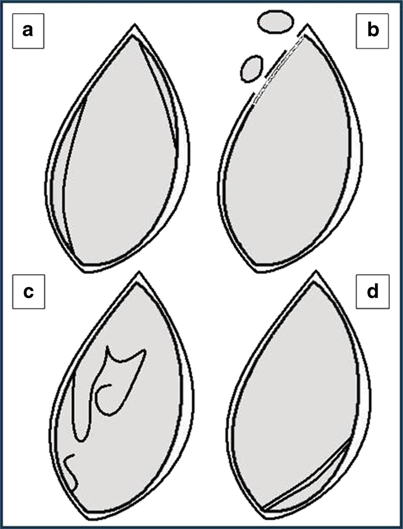
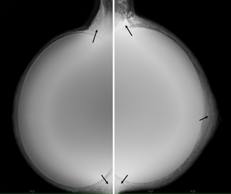
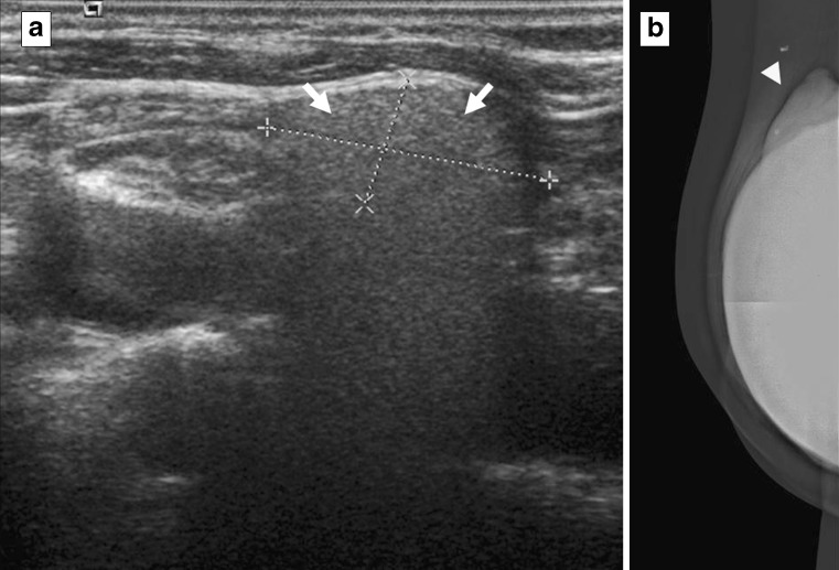
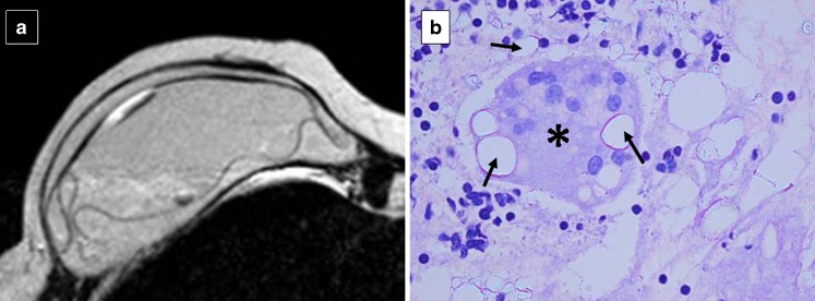
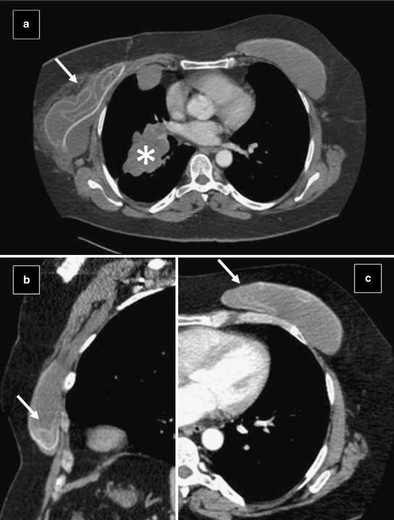
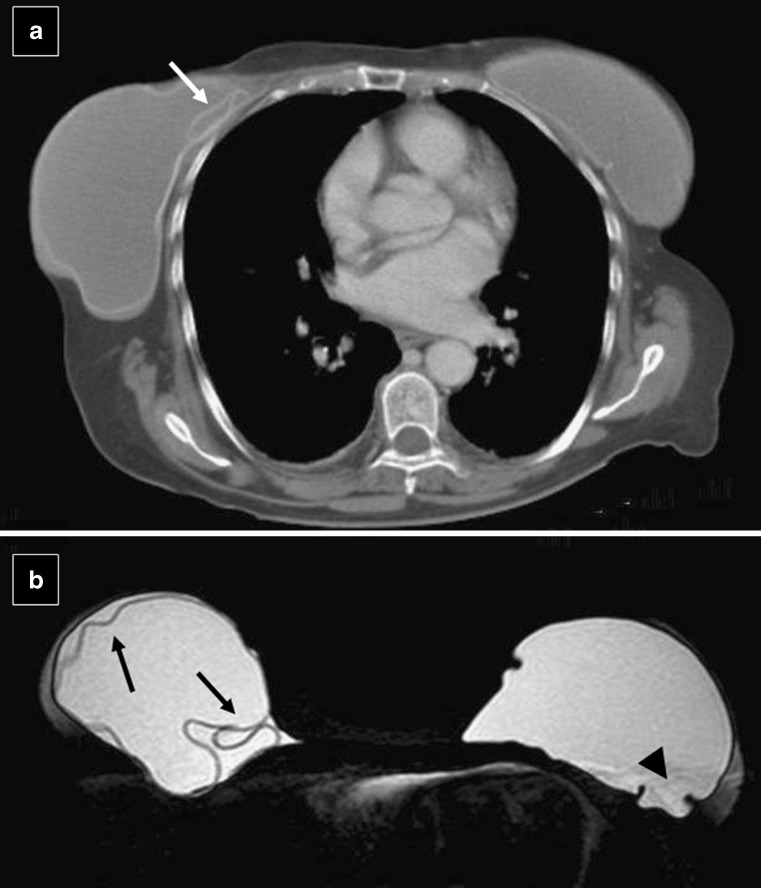
The number of women with breast implants is increasing. Radiologists must be familiar with the normal and abnormal findings of common implants. Implant rupture is a well-known complication after surgery and is the main cause of implant removal. Although mammography and ultrasonography are the standard first steps in the diagnostic workup, magnetic resonance imaging (MRI) is the most useful imaging modality for the characterisation of breast implants because of its high spatial resolution and contrast between implants and soft tissues and absence of ionising radiation. MRI has the highest sensitivity and specificity for implant rupture, thanks to its sequences that can suppress or emphasise the signal from silicone. Regardless of the technique used, the overall aim of imaging breast implants is to provide essential information about tissue and prosthesis integrity, detect implant abnormalities and detect breast diseases unrelated to implants, such as breast cancer.


Figures

Fig. 1
Fig. 1
Different implant types. a and b Breast tissue expander with metallic component visualised by computed tomography scan. Silicone gel implants with textured surface (c) and with smooth surface (d)
Fig. 2
Fig. 2
Localisation of breast implants. (Left) Submammary implant located in front of the pectoralis major muscle and (right) submuscular implant located behind the pectoralis major muscle, visualised by mammography
Fig. 3
Fig. 3
Magnetic resonance imaging scan of a woman with bilateral breast silicone implants. Right implant with extracapsular rupture exhibiting the typical “linguine sign” at the posterior margin of the implant. Intact left silicone implant
Fig. 4
Fig. 4
Our MRI examination protocol includes a 1.5-T superconducting MR system (Philips MR Systems Gyroscan NT) with a SENSE-body coil, with the following sequences. We always include a post-contrast study to detect possible malignant lesions
Fig. 5
Fig. 5
MRI of a 61-year-old woman with bilateral implants: a single-lumen implant (right breast) and a double-lumen implant (left breast). (a) Axial silicone-suppression and (b) axial T2-weighted turbo spin echo sequences. The right implant has homogeneous signal intensity, representing a single lumen with silicone gel (*). The left implant has an inner lumen (open arrow) of low-signal-intensity or high-signal-intensity silicone surrounded by a smaller outer lumen (solid arrow) that contains saline
Fig. 6
Fig. 6
Magnetic resonance imaging scan of a woman with bilateral ruptured implants. Typical “linguine sign” within implants representing collapsed implant shell
Fig. 7
Fig. 7
MRI of a woman with intracapsular rupture of a single-lumen silicone implant. (a) Axial T2-weighted turbo spin-echo and (b) axial silicone-excited sequence. The study shows a hypointense subcapsular line at the anterior margin of the implant (solid arrow); the “teardrop sign” and “key-hole sign” are also present (open arrows). Focal change in signal at the anterior margin of the implant (white open arrow) can also be observed
Fig. 8
Fig. 8
MRI of a woman with an extracapsular rupture of a single-lumen silicone implant. a and b Sagittal silicone-excited sequences demonstrate the presence of free silicone gel around the implant (white arrows). (c) Axial silicone-excited sequence shows free silicone gel located in the internal mammary chain (black arrow)
Fig. 9
Fig. 9
Axial T2-weighted turbo spin-echo image (a) and axial CT scan (c) of single-lumen implants show small amount of reactive fluid (arrows). (b) Axial T1-weighted turbo spin-echo image demonstrates normal radial folds of the membrane (arrowhead). Simple or complex folds are not in themselves indicative of rupture
Fig. 10
Fig. 10
Chest plain film of a woman with capsule calcification (arrowheads) adjacent to the implant. Many augmented patients develop capsular contracture
Fig. 11
Fig. 11
a Sagittal silicone-excited MRI sequence and (b) axial T2-weighted turbo spin-echo image of a 64-year-old woman with changes in the signal intensity of the silicone gel (black arrows). The margins of the implant are slightly irregular and a small amount of fluid surrounds the prosthesis (white arrow). A ruptured implant was confirmed at surgery.
Fig. 12
Fig. 12
MRI of a 54-year-old woman with a ruptured breast implant confirmed at surgery. a Axial silicone suppression. b Axial silicone-excited sequence. c Axial T2-weighted turbo spin echo. d Axial T1-weighted turbo spin echo. Silicone gel (white asterisks) inside and outside the implant. A moderate amount of water and probably serum is mixed in the silicone gel around the implant (black asterisks). Note also the punctuate changes in signal intensity—droplets within the implant (arrows) and punctuate and hyperintense images due to calcifications in the implant periphery (arrowheads)
Fig. 13
Fig. 13
Variants of normal breast implants. a Intact implant has an uninterrupted shell and fibrous capsule adjacent to the breast parenchyma. b Periprosthetic fluid. Presence of a small-to-moderate amount of reactive fluid surrounding the implant. c Simple or complex radial folds. Lines extending from the surface of the implant and inwards in a rather perpendicular manner. d Calcification and thickening of the fibrous capsule
Fig. 14
Fig. 14
Findings of possible breast implant rupture. a Deformity in contour. The border of the implant is bulging more than usual (called the “rat-tail sign” when very pronounced). Sometimes rupture cannot be differentiated from herniation. b Irregular margin. The border of the implant is blurry. Frequently seen with calcification of the fibrous capsule. c Changes in the signal intensity of the silicone gel. Water/serum mixed in the silicone gel through a defect in membrane. d “Noose sign” or “key-hole sign”. Small invagination of the shell where the two membranes do not touch. e “Teardrop sign”. Invagination of the shell containing a droplet of silicone. The last two images represent silicone gel leakage through a small focal implant shell tear
Fig. 15
Fig. 15
Definitive findings of breast implant rupture. a Subcapsular lines. Lines running almost parallel to the fibrous capsule and just beneath it. The beginning and the end of the line can be followed to the surface of the implant. b Siliconomas and free silicone. Disruption of the shell and fibrous capsule will allow silicone to extravasate into surrounding breast tissue. c “Linguine sign”. Folded wavy multidirectional lines within the silicone gel, representing the collapsed implant shell. d “Railroad track sign”. Two parallel lines in close proximity forming a double-contoured subcapsular line within the silicone gel
Fig. 16
Fig. 16
Oblique mammograms in a 29-year-old transsexual with subglandular implants and silicone injections. Diffuse areas of increased density are visualised adjacent to the implant (arrows)
Fig. 17
Fig. 17
Implant position is an important factor when studying the breasts. Patients with submammary implants have fewer visualised area compared to patients with submuscular implants. The displacement technique introduced by Eklund facilitates mammography in women with implants. Slightly more tissue is visualised with displacement (below) than with standard compression mammography (above)
Fig. 18
Fig. 18
a and b Ultrasonography of a woman with an intact implant. Breast gland (black asterisk), pectoralis major muscle (black arrow), and implant shell (white arrow) visualised as a thin and continuous echogenic line at the parenchymal tissue-implant interface, and silicone implant (white asterisk). (c) A small fluid collection around the implant (arrowhead) and (d) a simple infolding of the shell silicone implant (arrowhead)
Fig. 19
Fig. 19
(Above) A coronal maximum intensity projection from a silicone-excited sequence in a transsexual (Fig. 16) demonstrating multiple nodules with high signal throughout both breasts representing free silicone (arrows). (Below) It is extremely difficult to evaluate the silicone implants by ultrasonography because of attenuation of the ultrasound beam by the free injected silicone and granuloma formation in the subcutaneous tissue
Fig. 20
Fig. 20
a Coronal silicone-excited sequence and (b) coronal contrast-enhanced fat-suppressed T1-weighted image of the previous patient show multiple nodules of free silicone (“siliconomas”) in the gluteal muscles
Fig. 21
Fig. 21
Extracapsular silicone implant rupture in a 52-year-old woman with a history of breast cancer who presented with a palpable lesion in the supraclavicular right region. Mammogram shows an irregular lump from the implant (arrowhead) and ultrasonography demonstrates the presence of a nodular lesion with typical inhomogeneity (the “snowstorm sign”) at the posterior margin, suspicious for a lymph node containing silicone
Fig. 22
Fig. 22
Cytology of the node shows multinucleated foreign-body giant cells (arrowhead) with abundant birefringent particles inside and outside the cytoplasm, compatible with gel silicone (arrows). Axial T2-weighted turbo spin-echo MRI study corroborated an extracapsular rupture of the implant
Fig. 23
Fig. 23
Bilateral ruptured implants in a woman with primary lung cancer (asterisk). a Axial CT scan shows a severe deformity of the right implant surface representing a collapsed ruptured prosthesis (arrow). b Sagittal multiplanar reconstruction and (c) axial CT scan of the left implant show high-density curvilinear lines within the implant (“linguine sign”, arrows)
Fig. 24
Fig. 24
Unilateral implant rupture. (a) Axial CT scan shows small high-density lines within the silicone gel in the right implant, suggestive of collapsed rupture (arrow). (b) Axial silicone-excited MRI sequence confirmed intracapsular rupture, showing hypointense wavy lines at the posterior margin of the right implant (“linguine sign”) and subcapsular line at the anterior margin (arrows). Normal infoldings in the left implant (arrowhead)
Fig. 25
Fig. 25
Breast ultrasonography of a 39-year-old (a) and a 30-year-old (b) augmented women. In both studies there are two lesions (arrows) considered BIRADS III under follow-up. c Ultrasonography of a 46-year-old augmented woman with a history of breast cancer. US shows a lesion suspicious of recurrent tumor (arrow) that was confirmed by histology. Breast implant (asterisk)
Fig. 26
Fig. 26
An oblique mammogram of a 57-year-old woman with a submammary implant (a). A cluster of suspicious microcalcifications can be identified in the breast gland (arrows). A magnified mammogram (b) confirms the presence of malignant microcalcifications (arrows)
Fig. 27
Fig. 27
(Left) Metallic clip placed in the microcalcifications site after biopsy. (Right) Real-time ultrasound is used to guide the needle tip (arrow)

Similar articles

Cited by

References

    1. Frank S, Mahdi R, Sherko K. Imaging in patients with breast implants—results of the First International Breast (Implant) Conference 2009. Insights Imaging. 2010;1:93–97. doi: 10.1007/s13244-010-0021-z. - DOI - PMC - PubMed
    1. Yasuo A, Ritsu A, Shinichiro K, et al. Silicone-selective multishot echo-planar imaging for rapid MRI survey of breast implants. Eur Radiol. 2007;17:1875–1878. doi: 10.1007/s00330-006-0533-6. - DOI - PubMed
    1. Hölmich Lisbet R, Fryzek Jon P, Kim K, et al. The diagnosis of silicone breast-implant rupture clinical findings compared with findings at magnetic resonance imaging. Ann Plast Surg. 2005;54:583–589. doi: 10.1097/01.sap.0000164470.76432.4f. - DOI - PubMed
    1. Hölmich Lisbet R, Soren F, Fryzek Jon P, et al. Incidence of silicone breast implant rupture. Arch Surg. 2003;138:801–806. doi: 10.1001/archsurg.138.7.801. - DOI - PubMed
    1. Hölmich Lisbet R, Vejborg Ilse M, Carsten C, et al. The diagnosis of breast implant rupture: MRI findings compared with findings at explantation. Eur J Radiol. 2005;53:213–225. doi: 10.1016/j.ejrad.2004.03.012. - DOI - PubMed

LinkOut - more resources