. 2012;7(2):e30904.
doi: 10.1371/journal.pone.0030904. Epub 2012 Feb 13.

Enhanced functional recovery in MRL/MpJ mice after spinal cord dorsal hemisection

Affiliations

Enhanced functional recovery in MRL/MpJ mice after spinal cord dorsal hemisection

Sandrine Thuret et al. PLoS One. 2012.

Abstract

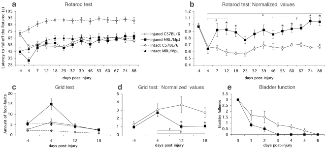
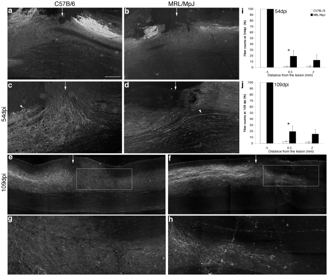
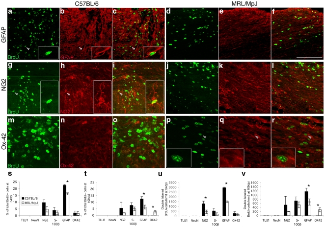
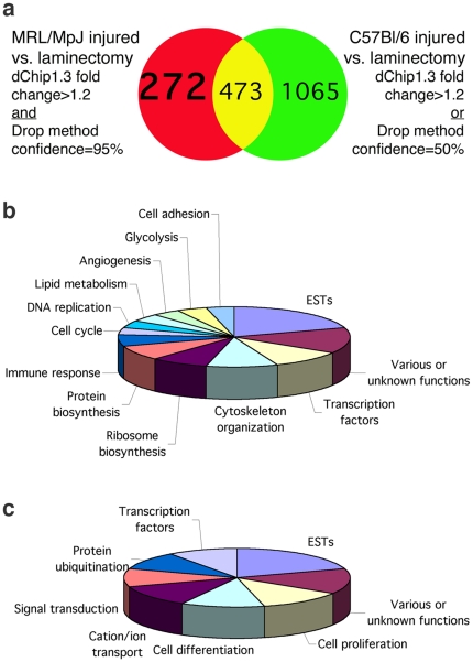
Adult MRL/MpJ mice have been shown to possess unique regeneration capabilities. They are able to heal an ear-punched hole or an injured heart with normal tissue architecture and without scar formation. Here we present functional and histological evidence for enhanced recovery following spinal cord injury (SCI) in MRL/MpJ mice. A control group (C57BL/6 mice) and MRL/MpJ mice underwent a dorsal hemisection at T9 (thoracic vertebra 9). Our data show that MRL/MpJ mice recovered motor function significantly faster and more completely. We observed enhanced regeneration of the corticospinal tract (CST). Furthermore, we observed a reduced astrocytic response and fewer micro-cavities at the injury site, which appear to create a more growth-permissive environment for the injured axons. Our data suggest that the reduced astrocytic response is in part due to a lower lesion-induced increase of cell proliferation post-SCI, and a reduced astrocytic differentiation of the proliferating cells. Interestingly, we also found an increased number of proliferating microglia, which could be involved in the MRL/MpJ spinal cord repair mechanisms. Finally, to evaluate the molecular basis of faster spinal cord repair, we examined the difference in gene expression changes in MRL/MpJ and C57BL/6 mice after SCI. Our microarray data support our histological findings and reveal a transcriptional profile associated with a more efficient spinal cord repair in MRL/MpJ mice.


Conflict of interest statement

Competing Interests: The authors have declared that no competing interests exist.

Figures

Figure 1
Figure 1. Enhanced functional recovery after SCI in MRL/MpJ mice.
a, The latency to fall off the rotarod is depicted as a function of time for intact (baseline) and injured MRL/MpJ and C57BL/6 mice. In both strains intact mice improved their performance over the course of the 88 days of training, but the C57BL/6 performed better at all time points tested. The injured MRL/MpJ reached their baseline level by the end of the 88 days of testing, whereas the injured C57BL/6 did not. b, Rotarod performance of injured mice is expressed as a ratio of baseline performance. Injured MRL/MpJ performed significantly better than injured C57BL/6 at 12, 18, 53, 60, 67, 74 and 88 dpi (*P<0.002). Injured MRL/MpJ significantly improved between 4 dpi and 88 dpi (#P<0.002), whereas injured C57BL/6 did not improve during that period. c, The number of foot-faults made during grid walking is reported as a function of time for intact (baseline) and injured MRL/MpJ and C57BL/6 mice. Both intact strains improved their ability to cross the grid with fewer foot-faults, but intact MRL/MpJ made more foot-faults throughout the entire training period. The injured MRL/MpJ reached their baseline level already by 12 dpi, whereas the injured C57BL/6 still had not reached their baseline level by the end of the testing period at 18 dpi. d, Grid walking performance of injured mice is expressed as a ratio to baseline performance. Injured MRL/MpJ performed significantly better than injured C57BL/6 at 12 and 18 dpi (p<0.05). Injured MRL/MpJ had significantly improved between 4 dpi and 12 dpi, 18 dpi (p<0.05), whereas injured C57BL/6 did not improve during that period. e, Estimation of bladder fullness is reported as a function of time after SCI in MRL/MpJ and C57BL/6 mice. All MRL/MpJ mice recovered autonomic bladder function by 3 dpi, whereas in C57BL/6 mice, autonomic bladder function did not returned until 6 dpi (Mann Whitney test, p-value = 0.028). Data present mean ± SEM. * denotes significant difference between MRL/MpJ and C57BL/6 and # denotes significant differences in MRL/MpJ performance between time points (P value: two-way ANOVA, Bonferroni post-hoc analysis).
Figure 2
Figure 2. Enhanced regeneration of corticospinal tract (CST) axons in MRL/MpJ mice after SCI.
a, b, Sagittal sections of BDA-labeled CST axons in C57BL/6 (a) and MRL/MpJ (b) mice. Two weeks after tracing, dorsal hemisections were performed. The CST was analyzed at 4 dpi ( = 18 days after tracing). In both mouse strains, axons are visible rostral and caudal from the injury site. No BDA-labeled fibers were found at the injury site, confirming that the CST was completely transected. c, d, Sagittal sections of BDA-labeled CST axons in injured C57BL/6 (c) and MRL/MpJ (d) mice at 54 dpi. Here, BDA tracing was performed right after SCI. C57BL/6 CST axons retracted from the lesion (arrowhead in c). In contrast, some MRL/MpJ CST fibers crossed through the lesion site (arrowhead in (d)). e, f, Montage of sagittal sections of BDA-labeled CST axons in C57BL/6 (e) and MRL/MpJ (f) mice at 109 dpi. Higher magnifications are shown in g (from boxed area in e) and h (from boxed area in f). We found no BDA-positive fibers below the injury site in C57BL/6 spinal cord (e, g), in contrast to MRL/MpJ mice (f, h). i, j, Data represent percentage of fibers and are reported as a function of the distance from the lesion at 54 dpi (i) and 109 dpi (j). MRL/MpJ mice exhibited significantly more regenerating axons at 0.5 mm caudal from the lesion site. Rostral end of the spinal cord is to the left. The arrows point at the injury site. Asterisks denote significant differences between MRL/MpJ and C57BL/6, P<0.02 (two-way ANOVA, Bonferroni Post-hoc). Scale bar, 250 µm (ad); 300 µm (e, f); 100 µm (g, h).
Figure 3
Figure 3. Reduced astrocytic response and micro-cavity formation in MRL/MpJ mice after SCI.
a, b, Sagittal sections of the lesion epicenter at 54 dpi labeled with GFAP (red) and counterstained with DAPI (blue) in C57BL/6 (a) and MRL/MpJ (b) mice. Higher magnifications are shown in c (from boxed area in a) and d (from boxed area in b). The dorsal surface of the injury site in the C57BL/6 mice bulged out and was filled with GFAP-positive astrocytes. In white and gray matter, numerous GFAP-positive astrocytes delineated the incision site (a, c). In MRL/MpJ mice, the dorsal surface of the injury site was concave and contained GFAP-negative cells surrounded by a few GFAP-positive astrocytes. The incision site was not visible any more (b, d). e, f, Sagittal sections of the lesion epicenter at 54 dpi stained for GFAP in C57BL/6 (e) and MRL/MpJ (f) mice. Numerous micro-cavities (arrowheads) are present at the injury site (arrows) in C57BL/6 mice (e) compared to none or few in MRL/MpJ mice (f). g, Bars represent the number of micro-cavities per cm3 at the injury epicenter at 54 dpi. C57BL/6 injured spinal cord developed significantly more micro-cavities than MRL/MpJ cord. h, i, Sagittal sections of the lesion epicenter at 109 dpi labeled with GFAP (red) and BrdU (green) in C57BL/6 (h) and MRL/MpJ (i) mice. The injury site of the C57BL/6 mice was filled with GFAP-positive astrocytes and micro-cavities were present (h). The injury site in the MRL/MpJ mice was not visible any more and could only be located by the presence of BrdU+ cells. The dorsal surface of the injury site was flat and few GFAP-positive astrocytes were present. (i). Asterisks denote significant difference between MRL/MpJ and C57BL/6, P<0.002 (Student's t-test). Scale bar, 250 µm (a, b, h, i); 140 µm (c, d); 125 µm (e, f).
Figure 4
Figure 4. Higher cell proliferation in intact spinal cord but lower increase in cell proliferation after SCI in MRL/MpJ mice.
a, b, Immunohistochemical staining for BrdU (arrowheads) on cross sections at the mid-thoracic level of intact spinal cord in C57BL/6 (a) and MRL/MpJ (b) mice 24 h after the last of 6 daily BrdU injections. c, Graphs show the amount of BrdU-labeled nuclei per mm3 in the intact spinal cord at the thoracic level 24 h and 4 weeks after the last BrdU injection. MRL/MpJ mice showed a 1.5× higher level of cell proliferation than C57BL/6. Cell survival was assessed at 4 weeks and there was no significant difference between the two strains. d, Number of BrdU-labeled nuclei per mm3 in the injured spinal cord 1, 54 and 109 dpi. BrdU was injected after injury and for the 6 following days. MRL/MpJ spinal cord showed less cell proliferation at 1 and 54 dpi. e, f, Sagittal sections of the lesion epicenter at 54 dpi stained for BrdU in C57BL/6 (e) and MRL/MpJ (f) mice. Asterisks denote significant difference between MRL/MpJ and C57BL/6, P<0.05 (Student's t-test). Scale bar, 260 µm.
Figure 5
Figure 5. Reduced astrocytic differentiation in MRL/MpJ injured spinal cord.
Sagittal sections of the lesion epicenter at 54 dpi (al) and 109 dpi (mr) labeled for BrdU (green) and lineage markers (red): GFAP (af), NG2 (gl), OX-42 (mr) in C57BL/6 (ac, gi, mo) and MRL/MpJ (df, jl, pr) mice. Higher magnifications of cells marked by the arrowheads are shown in boxed inserts. sv, Graphs represent the quantification of BrdU-labeled cells stained for a specific marker at 54 dpi (s, u) and 109 dpi (t, v) shown as percentage of all BrdU-labeled cells (s, t) and as absolute number of co-stained BrdU-labeled nuclei per mm3 (u, v). At 54 dpi, C57BL/6 mice showed more NG2/BrdU- and GFAP/BrdU-positive cells than MRL/MpJ mice. At 109 dpi, C57BL/6 mice had still more GFAP/BrdU-expressing cells than MRL/MpJ mice; however, OX-42/BrdU-positive cells were only found in MRL/MpJ spinal cord (2.7±0.9%). Asterisks denote significant difference between MRL/MpJ and C57BL/6, P<0.05 (Student's t-test). Scale bar, 100 µm (ai); 50 µm (pr); 50 µm for boxed area (ai); 25 µm for boxed area (pr).
Figure 6
Figure 6. Gene expression profiling of the MRL/MpJ and C57BL/6 injured spinal cord.
a, Using loose criteria (dChip1.3 fold change>1.2 OR Drop method confidence = 50%), 1538 genes were found to be differentially expressed in C57BL/6 spinal cord 4 days after hemisection. Using strict criteria (dChip1.3 fold change>1.2 AND Drop method confidence = 95%), 745 differentially expressed genes were found in MRL/MpJ spinal cord 4 days after hemisection, and 272 of these genes were exclusive for the MRL/MpJ injured spinal cord. b, Classification by biological function of genes up-regulated exclusively in MRL/MpJ injured spinal cord. c, Classification by biological function of genes down-regulated exclusively in MRL/MpJ injured spinal cord.

Similar articles

Cited by

References

    1. Thuret S, Moon LD, Gage FH. Therapeutic interventions after spinal cord injury. Nat Rev Neurosci. 2006;7:628–643. - PubMed
    1. Chen MS, Huber AB, van der Haar ME, Frank M, Schnell L, et al. Nogo-A is a myelin-associated neurite outgrowth inhibitor and an antigen for monoclonal antibody IN-1. Nature. 2000;403:434–439. - PubMed
    1. Fitch MT, Doller C, Combs CK, Landreth GE, Silver J. Cellular and molecular mechanisms of glial scarring and progressive cavitation: in vivo and in vitro analysis of inflammation-induced secondary injury after CNS trauma. J Neurosci. 1999;19:8182–8198. - PMC - PubMed
    1. Davies SJ, Fitch MT, Memberg SP, Hall AK, Raisman G, et al. Regeneration of adult axons in white matter tracts of the central nervous system. Nature. 1997;390:680–683. - PubMed
    1. Neumann S, Woolf CJ. Regeneration of dorsal column fibers into and beyond the lesion site following adult spinal cord injury. Neuron. 1999;23:83–91. - PubMed

Publication types