. 2012;7(2):e31809.
doi: 10.1371/journal.pone.0031809. Epub 2012 Feb 14.

SHARPIN is essential for cytokine production, NF-κB signaling, and induction of Th1 differentiation by dendritic cells

Affiliations

SHARPIN is essential for cytokine production, NF-κB signaling, and induction of Th1 differentiation by dendritic cells

Zhe Wang et al. PLoS One. 2012.

Abstract

Spontaneous mutations of the Sharpin (SHANK-associated RH domain-interacting protein, other aliases: Rbckl1, Sipl1) gene in mice result in systemic inflammation that is characterized by chronic proliferative dermatitis and dysregulated secretion of T helper1 (Th1) and Th2 cytokines. The cellular and molecular mechanisms underlying this inflammatory phenotype remain elusive. Dendritic cells may contribute to the initiation and progression of the phenotype of SHARPIN-deficient mice because of their pivotal role in innate and adaptive immunity. Here we show by flow cytometry that SHARPIN- deficiency did not alter the distribution of different DC subtypes in the spleen. In response to TOLL-like receptor (TLR) agonists LPS and poly I:C, cultured bone marrow-derived dendritic cells (BMDC) from WT and mutant mice exhibited similar increases in expression of co-stimulatory molecules CD40, CD80, and CD86. However, stimulated SHARPIN-deficient BMDC had reduced transcription and secretion of pro-inflammatory mediators IL6, IL12P70, GMCSF, and nitric oxide. Mutant BMDC had defective activation of NF-κB signaling, whereas the MAPK1/3 (ERK1/2) and MAPK11/12/13/14 (p38 MAP kinase isoforms) and TBK1 signaling pathways were intact. A mixed lymphocyte reaction showed that mutant BMDC only induced a weak Th1 immune response but stimulated increased Th2 cytokine production from allogeneic naïve CD4(+) T cells. In conclusion, loss of Sharpin in mice significantly affects the immune function of DC and this may partially account for the systemic inflammation and Th2-biased immune response.


Conflict of interest statement

Competing Interests: The authors have declared that no competing interests exist.

Figures

Figure 1
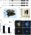
Figure 1. In vivo and in vitro features of SHARPIN.
(A) COILS and MotifScan programs were used to predict the presence of CC (coiled-coil) domain, UBL (ubiquitin-like) domain and ZFRBP (zinc-finger Ran-Binding protein 2) domain which form similar motif patterns in the SHARPIN protein of human, mouse and rat origins. (B) Eight week-old females of WT and cpdm mice. The mutant mice (above) develop progressive skin inflammation starting at about four weeks. (C) Extramedullary hematopoiesis causes marked enlargement of the spleen of cpdm mice. (D) BMDC were incubated in the absence or presence of 100 ng/ml LPS for 4 hours. The mRNA level of Sharpin was measured by qRT-PCR and presented relative to the mRNA expression in non-stimulated WT BMDC. Bars represent the mean ± s.d. of 3 mice. * P<0.05; ** P<0.001. (E) Fibroblast (NIH3T3) and macrophages (RAW264.7) cells were transfected with the expression plasmid pFLAG-SHARPIN. After 48 hours, cells were fixed and probed with anti-FLAG and FITC-conjugated secondary antibody. Nuclei were stained with DAPI. In both transfected cell lines, FLAG-SHARPIN was found to be cytoplasm-localized.
Figure 2
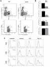
Figure 2. Effect of Sharpin mutation on DC subpopulations and maturation.
(A) Spleens from WT and cpdm mice were isolated and subject to collagenase and DNase digestion. The obtained splenic homogenates were centrifuged over a Percoll gradient (35% and 55% density) for 15 minutes. The bands at the 35%-medium and the 35–55% interface were pooled, washed and stained with a combination of various antibodies to stain different DC subsets, conventional CD11c+CD8α+, CD11c+CD8α and plasmacytoid DC (CD11cPDCA-1+).The top panels were gated on FSChiSSClo cells to show separate populations of CD11c+PDCA-1 and CD11cPDCA-1+ cells. Further gating on the CD11c+PDCA-1 subpopulation gave the bottom panel that showed two distinct pools of CD11c+CD8α+ and CD11c+CD8α cells. Percentages were calculated based on the parental population and were additionally shown as bar graphs (n = 2) (B). (C) WT and cpdm BMDC (5×10⁵) cells were stimulated with medium, 100 ng/ml LPS or 25 µg/ml poly I:C for 24 hours. The cells were labeled with PE-labeled anti-CD40, anti-CD80, and anti-CD86, and subjected to flow cytometry analysis. The populations shown in histograms were gated on CD11c+ cells. Unstained cells served as negative controls. Results are representative of two independent experiments.
Figure 3
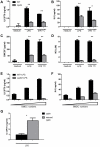
Figure 3. Defective production of pro-inflammatory mediators from stimulated cpdm BMDC.
(A–D) Cultured WT and cpdm BMDC (1×10⁵ cells in 0.1 ml complete medium) were washed and stimulated with medium, 100 ng/ml LPS or 25 µg/ml poly I:C for 24 hours. Supernatants were collected for ELISA of IL12P70, IL6, and GMCSF, and for quantification of nitric oxide (NO). (E–F) Gradient numbers (1×10⁴, 2×10⁴, and 4×10⁴ cells in 0.1 mL complete medium) of BMDC were used for100 ng/mL LPS stimulation. After 24 hours, the amounts of IL12P70 and IL6 from the supernatant were measured. (G) The cpdm mice rescued by Sharpin-containing BAC had complete remission of the inflammatory phenotype . BMDC developed from cpdm and rescued cpdm mice were plated (1×10⁶ cells in 0.3 mL) and stimulated with 100 ng/mL LPS. After 24 hours, supernatants were collected for analysis of IL12P70 production. Data are representative of three independent experiments. * P<0.01; ** P<0.005.
Figure 4
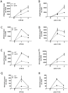
Figure 4. Decreased mRNA levels of inflammatory cytokines from cpdm BMDC.
Cultured WT and cpdm BMDC (5×10⁵ cells in 0.2 ml complete medium) were washed and stimulated with 100 ng/ml LPS (A,C,E,G) or 25 µg/ml poly I:C (B,D,F,H). At 0, 1 and 2 hours, total RNA was extracted and subject to qRT-PCR to measure the expression of Il12p40 (A,B), Il6 (C,D), Gmcsf (E,F), and Ifnb (G,H) mRNA. Bars represent mean ± SD. Data are representative of two independent experiments.
Figure 5
Figure 5. Normal TLR4 and MD2 expression and decreased IL10 secretion and A20 expression by cpdm BMDC.
(A) Unstimulated WT and cpdm BMDC (5×10⁵) were labeled with PE-labeled anti-TLR4/MD2 or anti-CD14 and then subject to flow cytometry analysis. Unstained cells serve as negative controls. (B) Cultured WT and cpdm BMDC (1×10⁵ cells in 0.1 ml complete medium) were washed and stimulated with medium, 100 ng/ml LPS or 25 µg/ml poly I:C for 24 hours. Supernatants were collected for ELISA of IL10. (C) Cultured WT and cpdm BMDC (5×10⁵ cells in 0.2 ml complete medium) were washed and stimulated with 100 ng/ml LPS or 25 µg/ml poly I:C. At 0, 1, and 2 hours, total RNA was extracted and subject to qRT-PCR to measure the production of A20.
Figure 6
Figure 6. Inhibition of NF-κB signaling in cpdm BMDC.
WT and cpdm BMDC (2×10⁶ cells in 0.5 mL complete medium) were stimulated with 100 ng/mL LPS (A) or 25 µg/mL poly I:C (B). At 0, 15, 30, and 60 minutes, whole-cell lysates were obtained and subject to immunoblots with antibodies against proteins involved in NF-κB, TBK1/IRF3, ERK1/2, and p38 signaling pathways. Beta-actin was used as loading control. (C) Cellular levels of p-IKK1/2 and p-IκBα in LPS- or poly I:C-stimulated BMDC were quantitated with ImageJ (NIH) and presented as trend lines. Results are representative of at least two independent experiments.
Figure 7
Figure 7. Stimulated cpdm BMDC induced Th2-biased cytokine production from naïve CD4+ T cells.
WT and cpdm BMDC (5×10⁴ cells in 0.1 mL complete medium; MHC haplotype: H-2b) were incubated with medium, 100 ng/mL LPS, 25 µg/mL poly I:C or 5 µg/mL Pam3CYS. After 24 hours, cells were washed with PBS and incubated with freshly isolated allogeneic naïve CD4+ T cells (2.5×10⁵ cells in 0.1 mL complete medium; MHC haplotype: H-2d). After 5 days, supernatants were collected and the secretion of IFNγ, IL4, and IL2 were measured by ELISA. Negative controls are 1) stimulated BMDC without co-culture with allogeneic CD4+ T cells; 2) allogeneic CD4+ T cells without co-culture with stimulated BMDC. Samples from both negative controls had no detectable production of the aforementioned cytokines (not shown). Results are analyzed based on 2–4 mice per group. * P<0.05.

Similar articles

Cited by

References

    1. Lim S, Sala C, Yoon J, Park S, Kuroda S, et al. Sharpin, a novel postsynaptic density protein that directly interacts with the shank family of proteins. Mol Cell Neurosci. 2001;17:385–397. - PubMed
    1. Seymour RE, Hasham MG, Cox GA, Shultz LD, Hogenesch H, et al. Spontaneous mutations in the mouse Sharpin gene result in multiorgan inflammation, immune system dysregulation and dermatitis. Genes Immun. 2007;8:416–421. - PubMed
    1. HogenEsch H, Gijbels MJ, Offerman E, van Hooft J, van Bekkum DW, et al. A spontaneous mutation characterized by chronic proliferative dermatitis in C57BL mice. Am J Pathol. 1993;143:972–982. - PMC - PubMed
    1. HogenEsch H, Janke S, Boggess D, Sundberg JP. Absence of Peyer's patches and abnormal lymphoid architecture in chronic proliferative dermatitis (cpdm/cpdm) mice. J Immunol. 1999;162:3890–3896. - PubMed
    1. HogenEsch H, Torregrosa SE, Boggess D, Sundberg BA, Carroll J, et al. Increased expression of type 2 cytokines in chronic proliferative dermatitis (cpdm) mutant mice and resolution of inflammation following treatment with IL-12. Eur J Immunol. 2001;31:734–742. - PubMed

Publication types