. 2012;7(2):e32030.
doi: 10.1371/journal.pone.0032030. Epub 2012 Feb 14.

Tumor susceptibility gene 101 (TSG101) is a novel binding-partner for the class II Rab11-FIPs

Affiliations

Tumor susceptibility gene 101 (TSG101) is a novel binding-partner for the class II Rab11-FIPs

Conor P Horgan et al. PLoS One. 2012.

Abstract

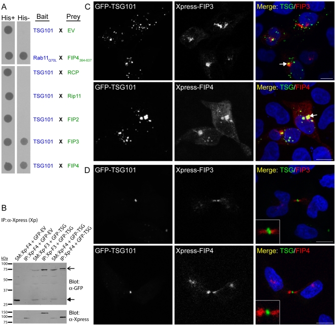
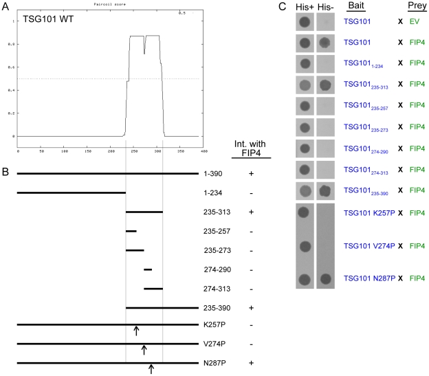
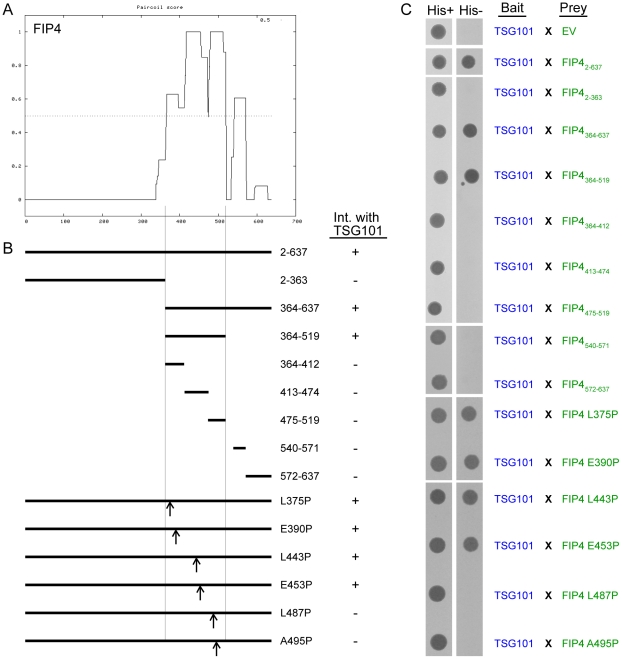
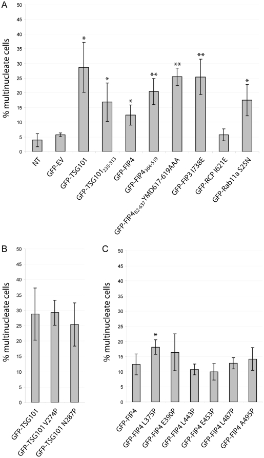
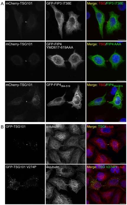
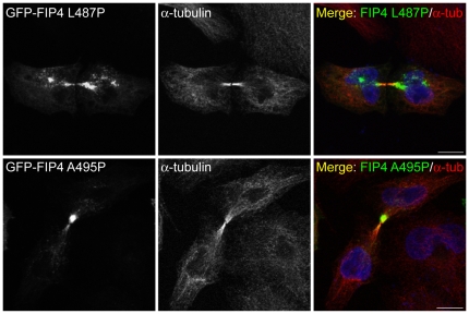
The Rab11-FIPs (Rab11-family interacting proteins; henceforth, FIPs) are a family of Rab11a/Rab11b/Rab25 GTPase effector proteins implicated in an assortment of intracellular trafficking processes. Through proteomic screening, we have identified TSG101 (tumor susceptibility gene 101), a component of the ESCRT-I (endosomal sorting complex required for transport) complex, as a novel FIP4-binding protein, which we find can also bind FIP3. We show that α-helical coiled-coil regions of both TSG101 and FIP4 mediate the interaction with the cognate protein, and that point mutations in the coiled-coil regions of both TSG101 and FIP4 abrogate the interaction. We find that expression of TSG101 and FIP4 mutants cause cytokinesis defects, but that the TSG101-FIP4 interaction is not required for localisation of TSG101 to the midbody/Flemming body during abscission. Together, these data suggest functional overlap between Rab11-controlled processes and components of the ESCRT pathway.


Conflict of interest statement

Competing Interests: The authors have declared that no competing interests exist.

Figures

Figure 1
Figure 1. TSG101 binds the class II FIPs.
(A) Yeast two-hybrid analysis of the interaction between the indicated proteins. Protein-protein interactions were determined by the ability of the transformed yeast to grow on minimal medium lacking tryptophan, leucine and histidine (His−). EV, empty vector. (B) Co-immunoprecipitation analysis of the ability of Xpress-FIPs to co-immunoprecipitate GFP-TSG101 in HeLa cells using an anti-Xpress antibody (SM, starting material; IP, immunoprecipitate). Co-immunoprecipitated proteins were revealed using an anti-GFP antibody. GFP-empty vector (EV) was used as a control. SM load was 3.33%. (C and D) HeLa cells were transfected with constructs encoding the indicated proteins. At 16–18 hours post-transfection, cells were processed for immunofluorescence microscopy and immunostained with an anti-Xpress antibody. Cells expressing relatively low levels of the GFP-TSG101 protein are shown in D. DAPI was used to visualise the nuclei. Images were acquired by confocal microscopy. Insets illustrate the midbody region of dividing cells at 2.5× higher magnification. Scale bar indicates 10 µm. Data are typical of at least three independent experiments.
Figure 2
Figure 2. The coiled-coil region of TSG101 mediates the interaction with FIP4.
(A) Plot depicting the probability of α-helical coiled-coil structure formation in TSG101 as determined using the PairCoil algorithm. (B) Schematic representation of the TSG101 truncation and point mutants that were tested for FIP4-binding ability. The outcome of the yeast two-hybrid experiments performed (part C) are indicated adjacent to the corresponding mutant in the schematic. (C) Yeast two-hybrid analysis of the interaction between the indicated proteins. Protein-protein interactions were determined by the ability of the transformed yeast to grow on minimal medium lacking tryptophan, leucine and histidine (His−). EV, empty vector. Data are typical of at least three independent experiments.
Figure 3
Figure 3. An extensive coiled-coil region of FIP4 mediates the interaction with TSG101.
(A) Plot depicting the probability of α-helical coiled-coil structure formation in FIP4 as determined using the PairCoil algorithm. (B) Schematic representation of the FIP4 truncation and point mutants that were tested for TSG101-binding ability. The outcome of the yeast two-hybrid experiments performed (part C) are indicated adjacent to the relevant mutant in the schematic. (C) Yeast two-hybrid analysis of the interaction between the indicated proteins. Protein-protein interactions were determined by the ability of the transformed yeast to grow on minimal medium lacking tryptophan, leucine and histidine (His−). EV, empty vector. Data are typical of at least three independent experiments.
Figure 4
Figure 4. FIP4 YMD617–619AAA is deficient in Rab11-binding.
HeLa cells were transfected with constructs encoding the indicated proteins. At 16–18 hours post-transfection, cells were processed for immunofluorescence microscopy and immunostained with an anti-Rab11a antibody. Images were acquired by confocal microscopy. Scale bar indicates 10 µm. Data are typical of at least three independent experiments.
Figure 5
Figure 5. Expression of TSG101 and FIP4 dominant-negative mutants cause abscission failure.
HeLa cells were transfected with constructs encoding the indicated proteins. At 36–40 hours post-transfection, cells were processed for immunofluorescence microscopy, immunostained for α-tubulin and their nuclei fluorescently-labelled with DAPI. A minimum of 150 transfected cells per experiment were counted and scored for multinucleation (>1 nucleus). Results, from three independent experiments, are expressed as the mean percentages ± S.D. Statistical significance was determined using an unpaired t test to investigate: (A) the difference between empty vector and GFP-fusion means, (B) the difference between GFP-TSG101 and GFP-TSG101 point mutant means and (C) the difference between GFP-FIP4 and GFP-FIP4 point mutant means. Statistical significance, *p<0.05, **p<0.02. NT, non-transfected; EV, empty vector.
Figure 6
Figure 6. TSG101 localises to the Flemming body during abscission independently of the class II FIPs.
(A and B) HeLa cells were transfected with constructs encoding the indicated proteins. At 16–18 hours post-transfection, cells were processed for immunofluorescence microscopy and, where indicated, immunostained for α-tubulin. DAPI was used to visualise the nuclei. Images, from cells expressing relatively low levels of the TSG101 fusion protein, were acquired by confocal microscopy. Scale bar indicates 10 µm. Data are typical of at least three independent experiments.
Figure 7
Figure 7. FIP4 localises to the midbody of dividing cells independently of TSG101.
HeLa cells were transfected with constructs encoding the indicated proteins. At 16–18 hours post-transfection, cells were processed for immunofluorescence microscopy and immunostained for α-tubulin. DAPI was used to visualise the nuclei. Images were acquired by confocal microscopy. Scale bar indicates 10 µm. Data are typical of at least three independent experiments.

References

    1. Glotzer M. The molecular requirements for cytokinesis. Science. 2005;307:1735–1739. - PubMed
    1. Glotzer M. Animal cell cytokinesis. Annu Rev Cell Dev Biol. 2001;17:351–386. - PubMed
    1. Horgan CP, McCaffrey MW. Endosomal trafficking in animal cytokinesis. Front Biosci (Schol Ed) 2012;4:547–555. - PubMed
    1. Raiborg C, Stenmark H. The ESCRT machinery in endosomal sorting of ubiquitylated membrane proteins. Nature. 2009;458:445–452. - PubMed
    1. Williams RL, Urbe S. The emerging shape of the ESCRT machinery. Nat Rev Mol Cell Biol. 2007;8:355–368. - PubMed

Publication types

MeSH terms