An encapsulated Yersinia pseudotuberculosis is a highly efficient vaccine against pneumonic plague
- PMID: 22348169
- PMCID: PMC3279354
- DOI: 10.1371/journal.pntd.0001528
An encapsulated Yersinia pseudotuberculosis is a highly efficient vaccine against pneumonic plague
Abstract
Background: Plague is still a public health problem in the world and is re-emerging, but no efficient vaccine is available. We previously reported that oral inoculation of a live attenuated Yersinia pseudotuberculosis, the recent ancestor of Yersinia pestis, provided protection against bubonic plague. However, the strain poorly protected against pneumonic plague, the most deadly and contagious form of the disease, and was not genetically defined.
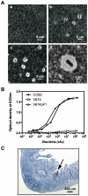
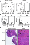
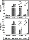
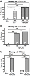
Methodology and principal findings: The sequenced Y. pseudotuberculosis IP32953 has been irreversibly attenuated by deletion of genes encoding three essential virulence factors. An encapsulated Y. pseudotuberculosis was generated by cloning the Y. pestis F1-encoding caf operon and expressing it in the attenuated strain. The new V674pF1 strain produced the F1 capsule in vitro and in vivo. Oral inoculation of V674pF1 allowed the colonization of the gut without lesions to Peyer's patches and the spleen. Vaccination induced both humoral and cellular components of immunity, at the systemic (IgG and Th1 cells) and the mucosal levels (IgA and Th17 cells). A single oral dose conferred 100% protection against a lethal pneumonic plague challenge (33×LD(50) of the fully virulent Y. pestis CO92 strain) and 94% against a high challenge dose (3,300×LD(50)). Both F1 and other Yersinia antigens were recognized and V674pF1 efficiently protected against a F1-negative Y. pestis.
Conclusions and significance: The encapsulated Y. pseudotuberculosis V674pF1 is an efficient live oral vaccine against pneumonic plague, and could be developed for mass vaccination in tropical endemic areas to control pneumonic plague transmission and mortality.
Conflict of interest statement
The authors have declared that no competing interests exist.
Figures


Similar articles
-
Complete Protection against Pneumonic and Bubonic Plague after a Single Oral Vaccination.PLoS Negl Trop Dis. 2015 Oct 16;9(10):e0004162. doi: 10.1371/journal.pntd.0004162. eCollection 2015. PLoS Negl Trop Dis. 2015. PMID: 26473734 Free PMC article.
-
Oral vaccination against plague using Yersinia pseudotuberculosis.Chem Biol Interact. 2017 Apr 1;267:89-95. doi: 10.1016/j.cbi.2016.03.030. Epub 2016 Apr 1. Chem Biol Interact. 2017. PMID: 27046452
-
A Recombinant Attenuated Yersinia pseudotuberculosis Vaccine Delivering a Y. pestis YopENt138-LcrV Fusion Elicits Broad Protection against Plague and Yersiniosis in Mice.Infect Immun. 2019 Sep 19;87(10):e00296-19. doi: 10.1128/IAI.00296-19. Print 2019 Oct. Infect Immun. 2019. PMID: 31331960 Free PMC article.
-
Current challenges in the development of vaccines for pneumonic plague.Expert Rev Vaccines. 2008 Mar;7(2):209-21. doi: 10.1586/14760584.7.2.209. Expert Rev Vaccines. 2008. PMID: 18324890 Free PMC article. Review.
-
Rational considerations about development of live attenuated Yersinia pestis vaccines.Curr Pharm Biotechnol. 2013;14(10):878-86. doi: 10.2174/1389201014666131226122243. Curr Pharm Biotechnol. 2013. PMID: 24372254 Free PMC article. Review.
Cited by
-
Plague Vaccine Development: Current Research and Future Trends.Front Immunol. 2016 Dec 14;7:602. doi: 10.3389/fimmu.2016.00602. eCollection 2016. Front Immunol. 2016. PMID: 28018363 Free PMC article. Review.
-
LcrV delivered via type III secretion system of live attenuated Yersinia pseudotuberculosis enhances immunogenicity against pneumonic plague.Infect Immun. 2014 Oct;82(10):4390-404. doi: 10.1128/IAI.02173-14. Epub 2014 Aug 11. Infect Immun. 2014. PMID: 25114109 Free PMC article.
-
The invasive pathogen Yersinia pestis disrupts host blood vasculature to spread and provoke hemorrhages.PLoS Negl Trop Dis. 2021 Oct 5;15(10):e0009832. doi: 10.1371/journal.pntd.0009832. eCollection 2021 Oct. PLoS Negl Trop Dis. 2021. PMID: 34610007 Free PMC article.
-
Common risk alleles for inflammatory diseases are targets of recent positive selection.Am J Hum Genet. 2013 Apr 4;92(4):517-29. doi: 10.1016/j.ajhg.2013.03.001. Epub 2013 Mar 21. Am J Hum Genet. 2013. PMID: 23522783 Free PMC article.
-
Selective Protective Potency of Yersinia pestis ΔnlpD Mutants.Acta Naturae. 2015 Jan-Mar;7(1):102-8. Acta Naturae. 2015. PMID: 25927007 Free PMC article.
References
-
- WHO. Human plague: review of regional morbidity and mortality, 2004–2009. Wkly Epidemiol Rec. 2009;85:40–45. - PubMed
-
- Arbaji A, Kharabsheh S, Al-Azab S, Al-Kayed M, Amr ZS, et al. A 12-case outbreak of pharyngeal plague following the consumption of camel meat, in north-eastern Jordan. Ann Trop Med Parasitol. 2005;99:789–793. - PubMed
-
- McClean KL. An outbreak of plague in northwestern province, Zambia. Clin Infect Dis. 1995;21:650–652. - PubMed
-
- Ratsitorahina M, Chanteau S, Rahalison L, Ratsifasoamanana L, Boisier P. Epidemiological and diagnostic aspects of the outbreak of pneumonic plague in Madagascar. Lancet. 2000;355:111–113. - PubMed
Publication types
MeSH terms
Substances
LinkOut - more resources
Full Text Sources
Other Literature Sources
Medical
Miscellaneous

