

Imaging molecular pathways: reporter genes
- PMID: 22348248
- PMCID: PMC3595018
- DOI: 10.1667/rr2918.1
Imaging molecular pathways: reporter genes
Abstract
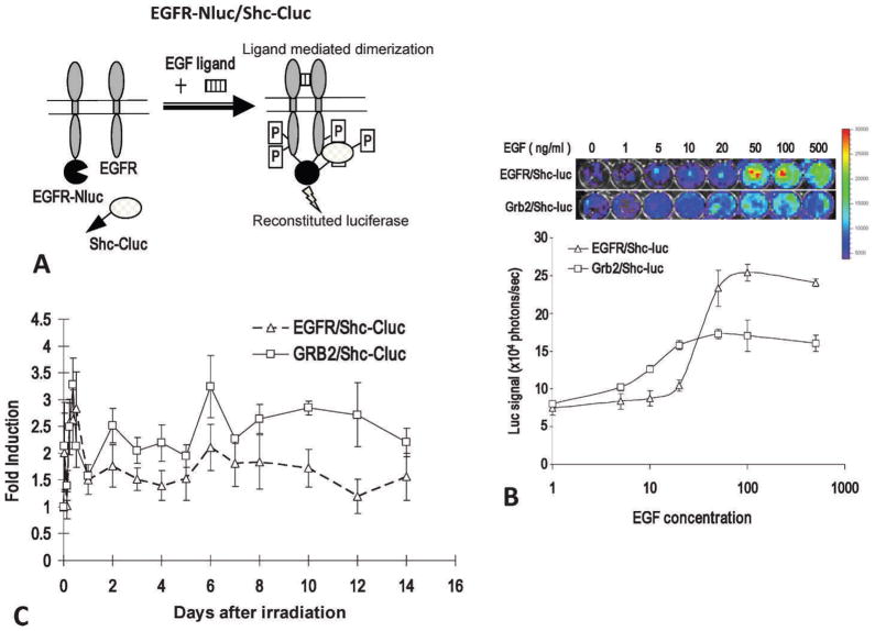
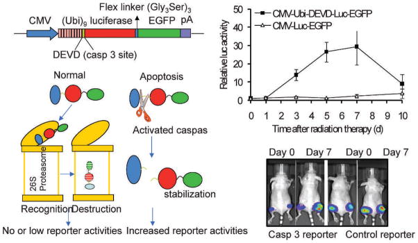
Molecular imaging is a rapidly advancing field that allows cancer biologists to look deeper into the complex inner workings of tumor cells, or whole tumors, in a non-invasive manner. In this review, we will summarize some recent advances that enable investigators to study various important biological processes in tumors in vivo. We will discuss novel imaging approaches that allow investigators to visualize and quantify molecular pathways, such as receptor tyrosine kinase activation, hypoxia signal transduction, apoptosis, and DNA double-strand breaks. Select examples of these applications will be discussed. Because of the limited scope of this review, we will only focus on natural reporters, such as bioluminescence and fluorescent proteins.
Figures
References
-
- Fowler AV, Zabin I. The amino acid sequence of beta galactosidase. I. Isolation and composition of tryptic peptides. J Biol Chem. 1970;245:5032–5041. - PubMed
-
- Shimomura O, Johnson FH, Saiga Y. Extraction, purification and properties of aequorin, a bioluminescent protein from the luminous hydromedusan, aequorea. J Cell Comp Physiol. 1962;59:223–239. - PubMed
-
- Prasher DC, Eckenrode VK, Ward WW, Prendergast FG, Cormier MJ. Primary structure of the Aequorea victoria green-fluorescent protein. Gene. 1992;111:229–233. - PubMed
-
- Chalfie M, Tu Y, Euskirchen G, Ward WW, Prasher DC. Green fluorescent protein as a marker for gene expression. Science. 1994;263:802–805. - PubMed
-
- Chudakov DM, Lukyanov S, Lukyanov KA. Fluorescent proteins as a toolkit for in vivo imaging. Trends Biotechnol. 2005;23:605–613. - PubMed
Publication types
MeSH terms
Substances
Grants and funding
LinkOut - more resources
Full Text Sources
Other Literature Sources
