

Oxidative stress imaging in live animals with techniques based on electron paramagnetic resonance
- PMID: 22348251
- PMCID: PMC3708477
- DOI: 10.1667/rr2668.1
Oxidative stress imaging in live animals with techniques based on electron paramagnetic resonance
Abstract
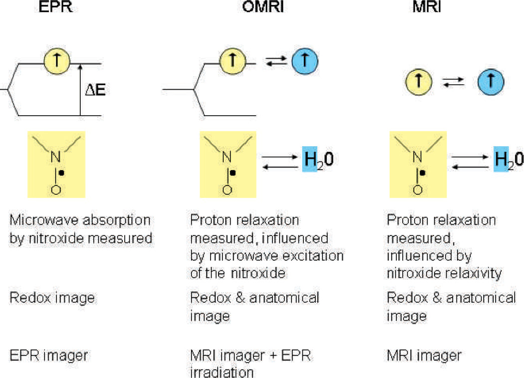
Oxidative stress has been the object of considerable biological and biochemical investigation. Quantification has been difficult although the quantitative level of products of biological oxidations in tissues and tissue products has emerged as a widely used technique. The relationship between these products and the amount of oxidative stress is less clear. Imaging oxidative stress with electron paramagnetic resonance related magnetic resonance imaging, while not addressing the specific issue of quantification of initiating events, focuses on the anatomic specific location of the oxidative stress. Moreover, the relative quantification of oxidative stress of one location against another is possible, sharpening our understanding of oxidative stress. This promises to improve our understanding of oxidative stress and its deleterious consequences and enhance our understanding of the effectiveness of interventions to modulate oxidative stress and its consequences.
Figures


References
-
- Martinovich GG, Martinovich IV, Cherenkevich SN, Sauer H. Redox buffer capacity of the cell: theoretical and experimental approach. Cell Biochem Biophys. 2010;58:75–83. - PubMed
-
- Griffiths HR, Moller L, Bartosz G, Bast A, Bertoni-Freddari C, Collins A, et al. Biomarkers. Mol Aspects Med. 2002;23:101–208. - PubMed
-
- Bartosz G. Use of spectroscopic probes for detection of reactive oxygen species. Clin Chim Acta. 2006;368:53–76. - PubMed
-
- Wang H, Joseph JA. Quantifying cellular oxidative stress by dichlorofluorescein assay using microplate reader. Free Radic Biol Med. 1999;27:612–616. - PubMed
-
- Wardman P. Fluorescent and luminescent probes for measurement of oxidative and nitrosative species in cells and tissues: progress, pitfalls, and prospects. Free Radic Biol Med. 2007;43:995–1022. - PubMed
Publication types
MeSH terms
Substances
Grants and funding
LinkOut - more resources
Full Text Sources
