Review
. 2012 Sep;16(9):1978-90.
doi: 10.1111/j.1582-4934.2012.01552.x.

Inflammation in atherosclerosis: a cause or a result of vascular disorders?

Affiliations
Review

Inflammation in atherosclerosis: a cause or a result of vascular disorders?

Ileana Manduteanu et al. J Cell Mol Med. 2012 Sep.

Abstract

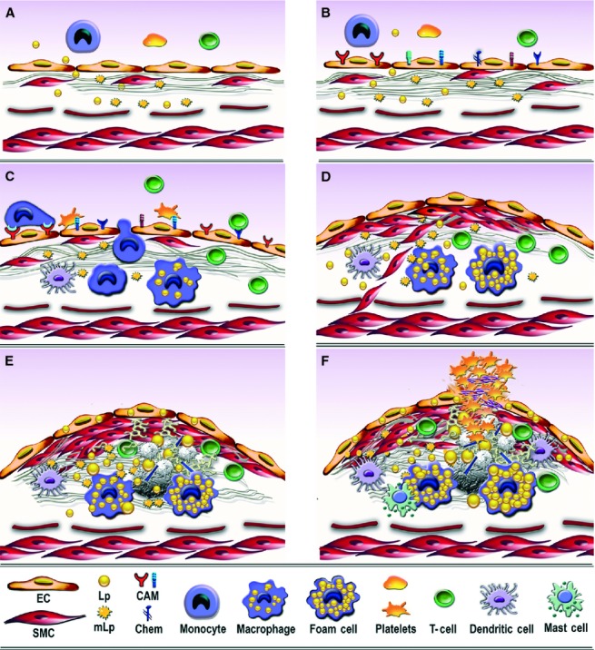
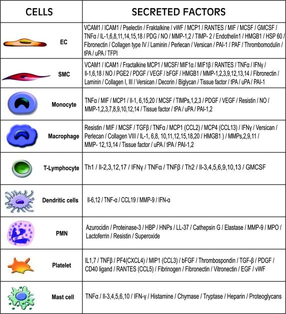
Sound data support the concept that in atherosclerosis, inflammation and dyslipidemia intersect each other and that irrespective of the initiator, both participate from the early stages to the ultimate fate of the atheromatous plaque. The two partakers manoeuvre a vicious circle in atheroma formation: dyslipidaemia triggers an inflammatory process and inflammation elicits dyslipidaemia. Independent of the initial cause, the atherosclerotic lesions occur focally, in particular arterial-susceptible sites, by a process that, although continuous, can be arbitrarily divided into a sequence of consecutive stages that lead from fatty streak to the fibro-lipid plaque and ultimately to plaque rupture and thrombosis. In the process, the initial event is a change in endothelial cells (EC) constitutive properties. Then, the molecular alarm signals send by dysfunctional EC are decoded by specific blood immune cells (monocytes, T lymphocytes, neutrophils, mast cells) and by the resident vascular cells, that respond by initiating a robust inflammatory process, in which the cells and the factors they secrete hasten the atheroma development. Direct and indirect crosstalk between the cells housed within the nascent plaque, complemented by the increase in risk factors of atherosclerosis lead to atheroma development and outcome. The initial inflammatory response can be regarded as a defense/protective reaction mechanism, but its further amplification, speeds up atherosclerosis. In this review, we provide an overview on the role of inflammation and dyslipidaemia and their intersection in atherogenesis. The data may add to the foundation of a novel attitude in the diagnosis and treatment of atherosclerosis.


Figures

Fig 1
Fig 1
Consecutive arbitrary stages occurring in the development of atherosclerotic lesion in arterial lesion-prone areas. (A) Stage I. Endothelial cell activation/modulation of constitutive functions. The initial stage in atheroma formation in dyslipidaemia consists in endothelial cell (EC) increased transcytosis of plasma lipoproteins (Lp) and their housing in the subendothelium and a switch of the cells to a secretory phenotype responsible for the development of a hyperplasic basal lamina. Within the subendothelium, Lp, especially LDL by interaction with extracellular matrix components, changes its attributes becoming the highly atherogenic, oxidized modified lipoproteins (mLp). (B) Stage II. EC dysfunction. Affected on both sides, luminal by alterations of plasma homoeostasis and abluminal by the accrual of mLp, the EC initiate an inflammatory process manifested by the expression of new or more cell adhesion molecules, cytokines and chemokines (CAM and Chem), an indication of endothelial dysfunction. (C) Stage III. Recruitment of blood immune cells and commencement of a robust inflammatory reaction. Blood monocytes and T cells adhere to activated/dysfunctional EC and undergo directed diapedesis into the intima. Adhered platelets assist leucocytes migration. Within the intima, monocytes become activated macrophages that express scavenger receptors, which function in the uptake of mLp and the formation of foam cells that secrete a variety of proinflammatory mediators. Lymphocytes switch to activated pro-inflammatory (Th 1) and anti-inflammatory (Th 2 and TREG) cells that secrete cytokines and chemokines. The direct or indirect crosstalk between resident and migrated cells within the intima dictates the lesion progression. Activated dendritic cells contribute to T cells recruitment and activation within the plaque. (D) Stage IV. SMC-key participants to fibrous plaque formation. The proliferation of intima-resident SMC and of SMC migrated from the media to the intima leads to the formation of a fibrous cap that is accompanied by increased synthesis of extracellular matrix components. (E) Stage V. Resident and immune cells and the factors they secrete generate a calcified fibro-lipid plaque. SMC-, macrophages-derived foam cells, apoptotic cells-derived lipids and calcification centres form a lipid loaded necrotic core rich in cholesterol crystals. (F) Stage VI. The unstable fibro-lipid plaque: rupture and thrombosis EC apoptosis and erosion, thinning of the fibrous cap, cell apoptosis, macrophages, dendritic and mast cells-secreted pro-inflammatory mediators generate the physical rupture of the plaque. This ends in direct contact between tissue factors and circulating platelets and blood coagulation components triggering the thrombus formation that may partially or totally imped the blood flow leading to either myocardial infarction or stroke.
Fig 2
Fig 2
Ultrastructural evidence of the initial endothelial cell (EC) changes occurring in hamster atheroma formation. (A) Accumulation beneath the activated EC of modified lipoproteins (mLp) under and within the basal lamina (bl). Bar: 0.1 μm. The mLp appear as a heterogeneous population of vesiculated, aggregated, or fused particle (arrows). (B) Hyperplasia of EC basal lamina (bl) as a result of the switch of the endothelium to a secretory phenotype; the bl appears in multiple interconnected rows. Bar: 0.1 μm.
Fig 3
Fig 3
Diagrammatic representation of the implicated vascular resident and recruited cells and the factors they secrete in the course of atheroma development.
Fig 4
Fig 4
Ultrastructural evidence of the cross-talk between an endothelial cell (EC), a T cell and a monocytes-derived foam cell (mFC) located in an area of subendothelial accumulation and retention of modified lipoproteins (mLp) in a hypercholesterolaemic hamster valve. Note the direct contact between the EC and the T cell and via pseudopodes between EC and mFC (arrows) and between mFC and T cell (arrow head). l, vascular lumen. ×14,000.

Similar articles

Cited by

References

    1. Libby P, Ridker PM, Hansson GK. Inflammation in atherosclerosis: from pathophysiology to practice. J Am Coll Cardiol. 2009;54:2129–38. - PMC - PubMed
    1. Millonig G, Malcom GT, Wick G. Early inflammatory-immunological lesions in juvenile atherosclerosis from the pathobiological determinants of atherosclerosis in youth (PDAY)-study. Atherosclerosis. 2002;160:441–8. - PubMed
    1. Tsimikas S, Willerson JT, Ridker PM. C-reactive protein and other emerging blood biomarkers to optimize risk stratification of vulnerable patients. J Am Coll Cardiol. 2006;47:C19–31. - PubMed
    1. Simionescu N, Vasile E, Lupu F, et al. Prelesional events in atherogenesis: accumulation of extracellular cholesterol rich liposomes in the arterial intima and cardiac valves of hyperlipidemic rabbits. Am J Pathol. 1986;123:85–101. - PMC - PubMed
    1. VanderLaan PA, Reardon CA, Getz GS. Site-specificity of atherosclerosis: site-selective responses to atherosclerotic modulators. Arterioscler Thromb Vasc Biol. 2004;24:12–22. - PubMed

Publication types

MeSH terms