. 2012 May 1;52(9):1692-7.
doi: 10.1016/j.freeradbiomed.2012.02.015. Epub 2012 Feb 17.

A mutant light-chain ferritin that causes neurodegeneration has enhanced propensity toward oxidative damage

Affiliations

A mutant light-chain ferritin that causes neurodegeneration has enhanced propensity toward oxidative damage

Martin A Baraibar et al. Free Radic Biol Med. .

Abstract

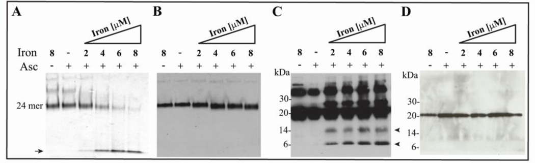
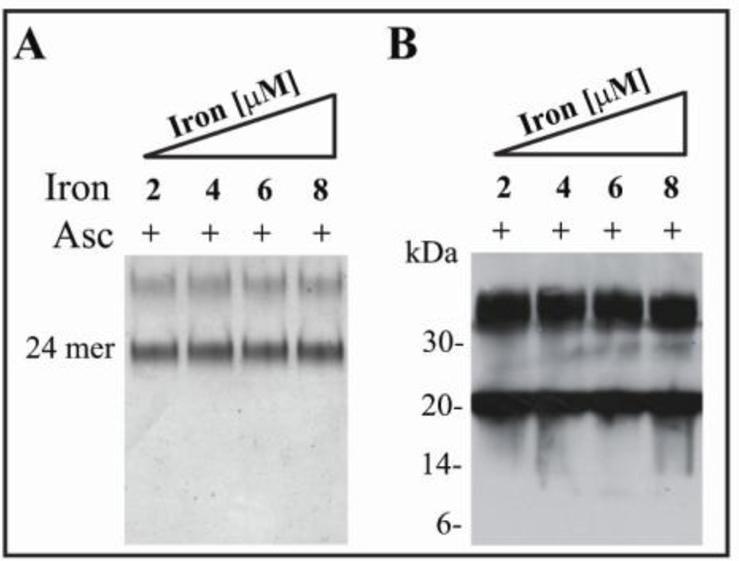
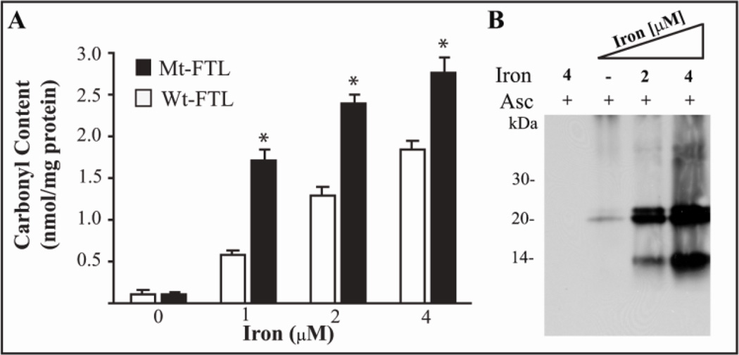
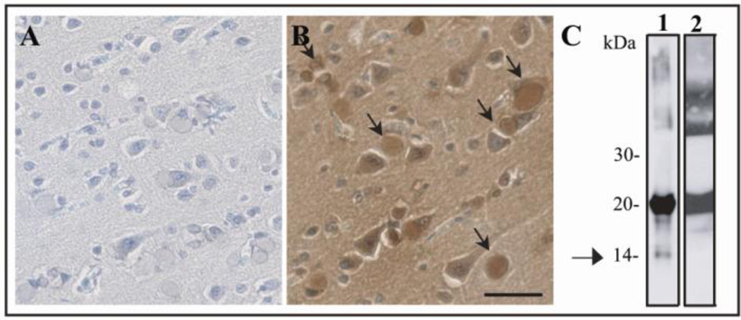
Intracellular inclusion bodies (IBs) containing ferritin and iron are hallmarks of hereditary ferritinopathy (HF). This neurodegenerative disease is caused by mutations in the coding sequence of the ferritin light chain (FTL) gene that generate FTL polypeptides with a C-terminus that is altered in amino acid sequence and length. Previous studies of ferritin formed with p.Phe167SerfsX26 mutant FTL (Mt-FTL) subunits found disordered 4-fold pores, iron mishandling, and proaggregative behavior, as well as a general increase in cellular oxidative stress when expressed in vivo. Herein, we demonstrate that Mt-FTL is also a target of iron-catalyzed oxidative damage in vitro and in vivo. Incubation of recombinant Mt-FTL ferritin with physiological concentrations of iron and ascorbate resulted in shell structural disruption and polypeptide cleavage not seen with the wild type, as well as a 2.5-fold increase in carbonyl group formation. However, Mt-FTL shell disruption and polypeptide cleavage were completely inhibited by the addition of the radical trap 5,5-dimethyl-1-pyrroline N-oxide. These results indicate an enhanced propensity of Mt-FTL toward free radical-induced oxidative damage in vitro. We also found evidence of extensive carbonylation in IBs from a patient with HF together with isolation of a C-terminal Mt-FTL fragment, which are both indicative of oxidative ferritin damage in vivo. Our data demonstrate an enhanced propensity of mutant ferritin to undergo iron-catalyzed oxidative damage and support this as a mechanism causing disruption of ferritin structure and iron mishandling that contribute to the pathology of HF.


Figures

Figure 1
Figure 1. Shell disruption and polypeptide cleavage of recombinant Mt-FTL homopolymers in the presence of iron and ascorbate
Wt- and Mt-FTL apoferritin homopolymers (0.5 µM) were incubated with increasing concentrations of ferrous ammonium sulphate (2, 4, 6 and 8 µM) in Hepes buffer (100 mM, pH 7) in the presence of ascorbate (2 mM) for 1 h in the dark at 24°C. Homopolymers (0.5 µM) were also incubated with iron (8 µM) but without ascorbate (lane 1), and with ascorbate (2 mM) without iron (lane 2). Native-PAGE of Mt-FTL (A) and Wt-FTL (B) homopolymers stained with Coomasie blue. Western blot analysis of Mt-FTL homopolymers (C) and Wt-FTL homopolymers (D) after SDS-PAGE using an antibody specific for the C-terminus of Mt-and Wt-FTL (23).
Figure 2
Figure 2. Prevention of shell disruption and polypeptide cleavage of recombinant Mt- FTL homopolymers by DMPO
Mt-FTL apoferritin homopolymers (0.5 µM) were incubated with increasing concentrations of iron (2, 4, 6 and 8 µM) in the presence of ascorbate (2 mM) and DMPO (50 mM). Samples were electrophoresed with native PAGE and stained with Coomasie blue (A) or in SDS-PAGE and blotted using an antibody specific for the C-terminus of Mt-FTL (B).
Figure 3
Figure 3. Protein carbonyl group formation in recombinant ferritin by treatment with iron and ascorbate
Wt- and Mt-FTL apoferritin homopolymers (0.5 µM) were incubated with increasing concentrations of iron (0, 1, 2, and 4 µM) in the presence of ascorbate (2 mM) and then derivatized with DNP-H. Carbonyl content was quantified by ELISA. Asterisks indicate statistically significant differences (p<0.05) between Mt- and Wt-FTL carbonyls content (A). Carbonyl groups were detected on Mt-FTL samples by western blot after SDS-PAGE using an antibody specific for DNP As controls, recombinant ferritins were incubated with iron (4 uM) and ascorbate (2 mM) but not treated with DNPH (lane 1) (B).
Figure 4
Figure 4. Protein carbonylation and Mt-FTL polypeptide cleavage in IBs from a HF patient carrying the c.497_498dupTC mutation
Immunohistochemistry for carbonyl group detection was performed in post-mortem brain tissue from a patient with HF, in which sections were not treated (A) or treated (B) with DNP-H. Carbonyl immunoreactivity was found co-localized with ferritin IBs (arrows). Bar: 25 µm. Western blot analysis (C) after SDS-PAGE of insoluble (lane 1) and soluble (lane 2) fractions obtained from IBs isolated from the putamen of the same patient blotted using an antibody specific for the C-terminus of Mt-FTL (23). The arrow indicates the presence of an ~14 kDa Mt-FTL C-terminal fragment visible only in the insoluble fraction.

References

    1. Stadtman ER, Berlett BS. J Biol Chem. 1991;266:17201–17211. - PubMed
    1. Fenton HJH. J Chem Soc Trans. 1896;69:546–562.
    1. Haber F, Weiss J. Naturwiss. 1932;51:948–950.
    1. Stadtman ER. Free Radical Biol. Med. 1990;9:315–325. - PubMed
    1. Stadtman ER, Berlett BS. Drug Metab Rev. 1998;30:225–243. - PubMed

Publication types

MeSH terms