Review
. 2012 Aug;69(15):2469-83.
doi: 10.1007/s00018-012-0928-2. Epub 2012 Feb 15.

Cytoskeleton responses in wound repair

Affiliations
Review

Cytoskeleton responses in wound repair

Maria Teresa Abreu-Blanco et al. Cell Mol Life Sci. 2012 Aug.

Abstract

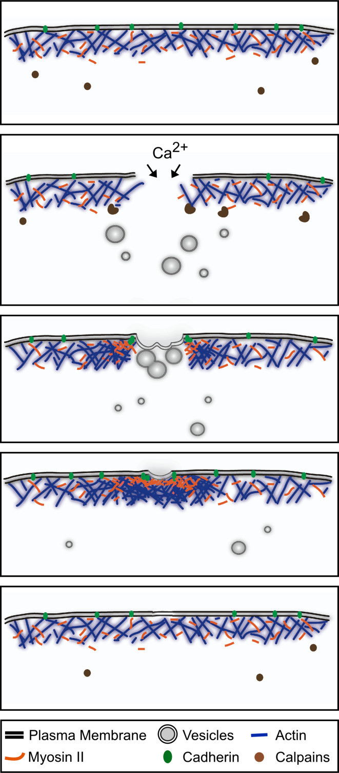
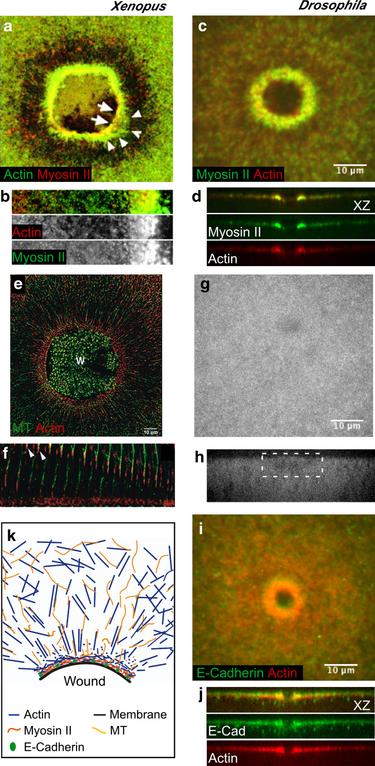
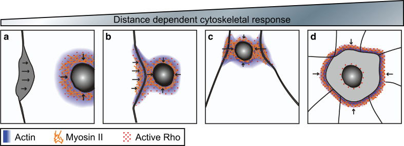
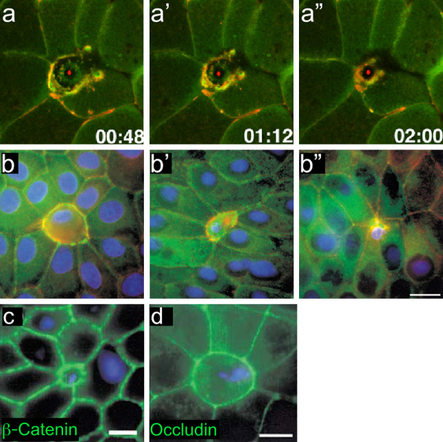
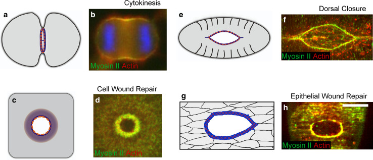
Wound repair on the cellular and multicellular levels is essential to the survival of complex organisms. In order to avoid further damage, prevent infection, and restore normal function, cells and tissues must rapidly seal and remodel the wounded area. The cytoskeleton is an important component of wound repair in that it is needed for actomyosin contraction, recruitment of repair machineries, and cell migration. Recent use of model systems and high-resolution microscopy has provided new insight into molecular aspects of the cytoskeletal response during wound repair. Here we discuss the role of the cytoskeleton in single-cell, embryonic, and adult repair, as well as the striking resemblance of these processes to normal developmental events and many diseases.


Figures

Fig. 1
Fig. 1
Single cell wound repair. Schematic diagram of the single-cell wound-repair process depicting the plasma membrane and cytoskeleton components. Upon plasma membrane disruption, Ca2+ influx and other signals trigger the temporal disassembly of the cortical cytoskeleton. Simultaneously, internal vesicles fuse with each other to form a membrane patch that will directionally migrate to the wound site and fuse with the plasma membrane. Once the membrane has been resealed, the plasma membrane and cortical cytoskeleton are remodeled. This process involves the assembly of an actomyosin ring, the replacement of the membrane patch, and the reconstitution of the cortical cytoskeleton
Fig. 2
Fig. 2
Cytoskeleton components of the single cell wound repair response. Fluorescent micrographs of single cell wound repair in Xenopus oocytes (a, b, e, f) and the Drosophila embryo (c, d, gj), showing the dynamic response of different cytoskeletal components upon wounding. ad An actomyosin contractile array is assembled at the wound edge a few seconds post-wounding (30–45 s). Actin and/or myosin-enriched zones can be distinguished: myosin II is enriched in the leading edge of the wound followed by an overlapping actin-myosin-enriched zone then a broad array of actin (b, d) (a, b adapted from [18], originally published in Journal of Cell Biology. doi:10.1083/jcb.200103105) (c, d adapted from [13], originally published in Journal of Cell Biology. doi:10.1083/jcb.201011018). ef Cell wounding triggers the formation of a radial arrangement of microtubules in Xenopus oocytes. Microtubules are transported to the wound in association with actin (arrowheads in f), and modulate the assembly of the actomyosin array (adapted from [19], and reproduced with permission from Elsevier). g, h In the Drosophila embryo, no radial rearrangement of the microtubule network is observed upon wounding (wound site is indicated by white box) (adapted from [13], originally published in Journal of Cell Biology. doi:10.1083/jcb.201011018). i, j E-Cadherin co-localizes with the actomyosin array in single cell wounds in Drosophila, and tethers the ring to the plasma membrane (adapted from [13], originally published in Journal of Cell Biology. doi:10.1083/jcb.201011018). k Cartoon depicting the response of the different cortical cytoskeleton components to cell wounding (adapted from [12])
Fig. 3
Fig. 3
Distance-dependent cytoskeletal responses triggered by a damaged single cell within an epithelia. Arrows indicate direction of repair forces. ad Cartoon depicting the response of neighboring cells upon wounding and the formation of a hybrid purse string in Xenopus oocytes. Wounds distal from neighboring cells repair via formation of an actomyosin purse string, but detach from neighbors as the wound closes (a), while wounds made near to neighboring cells form a hybrid purse string where the actomyosin contractile array forms at the wound as well as at nearby cell contacts (b), as the wound closes the cell contacts ingress (c). Severely damaged cells are extruded from the epithelia by the formation of an actomyosin contractile array between the surrounding neighbor cells (d)
Fig. 4
Fig. 4
Cells unable to undergo repair in a multicellular context resemble cell extrusion during apoptosis. aa″ Time-lapse images of a single cell being removed from the Xenopus embryo epithelia. The apoptotic cell is extruded by an actomyosin ring assembling between neighboring cells (adapted from [24], and reproduced with permission from Elsevier). bb″ Time series showing the interface between an apoptotic cell and its neighbors at early (b), middle (b′), and late (b″) stages of apoptotic cell extrusion. Staining for myosin II (green), actin (red), and DNA (blue) shows that actin and myosin II co-localize around the apoptotic cell. c, d β-catenin (c) and occludin (d) staining shows that adherens and tight junctions are maintained between the apoptotic cell and surrounding tissue, indicating that tissue integrity is maintained during the extrusion process (adapted from [26], and reproduced with permission from Elsevier)
Fig. 5
Fig. 5
Mechanisms of embryo epithelial repair. aa′″ Cartoon depicting the cell rearrangements and the assembly of the actomyosin purse string in epithelial wound repair. The actomyosin purse string is intercellularly linked through E-cadherin-based adherens junctions. Arrows indicate the cells that will be removed from the leading edge and moved back during repair. bb′″ Confocal series showing actin protrusions at the wound leading edge. These protrusions use each other as tethers to close the wound (adapted from [31], and reproduced with permission from Nature Publishing Group). cc′ In mouse embryonic epithelia, the mesenchyme is exposed upon wounding, and its contraction mediates wound closure. Mesenchyme contraction was followed with DiI for 0 h (c) and 12 h (c′) post-wounding (adapted from [91], and reproduced with permission from Springer). dd′ Cartoon depicting the components contributing to epithelial wound repair: apical layer cell migration and contraction of the underlying mesenchyme (adapted from [91])
Fig. 6
Fig. 6
Adult epithelial repair. Three to 10 days after the initial wound, new tissue is being formed and remodeled in the wound bed. The initial clot is slowly sloughed off as a scab, while the underlying keratinocytes migrate to re-epithelialize the wound. As the keratinocytes migrate, they deposit a new basement membrane and undergo rapid cycles of cell division in order to completely restore the epidermis. At this stage, fibroblasts and new capillaries have populated the underlying dermis. The fibroblasts have begun to differentiate into proto-myofibroblasts and eventually myofibroblasts, which will continuously replace the fibers that make up the ECM and apply powerful contractile forces to the wound bed
Fig. 7
Fig. 7
Keratinocytes in wound repair. a, b During wound repair, keratinocytes shift from a stationary barrier into highly motile invasive cells requiring dramatic cytoskeletal reconfiguration. Cartoon depicting a stationary keratinocyte in healthy epithelia (a) and migrating through a wound bed (b). c Spreading human primary keratinocyte stained for the focal adhesion protein vinculin (green), F-actin (red) and the nucleus (blue) (adapted from The Cell: An Image Library, CIL:12655, contributed by Julien Gautrot)
Fig. 8
Fig. 8
Fibroblast differentiation and activity in the wound bed. Fibroblasts differentiate into proto-myofibroblasts and eventually into myofibroblasts as they contract the wound bed and remodel the ECM. a Cartoon depicting the transformations of the cell’s cytoskeleton and the surrounding ECM as the fibroblast differentiates into a myofibroblast (adapted from [38], with permission from Nature Publishing Group). b Rat subcutaneous fibroblasts were differentiated into myofibroblasts in vitro. Cells were immunostained for αSMA (red) to visualize contractile stress fibers, F-actin (green), and nuclei (blue) (adapted from [92], with permission from Nature Publishing Group)
Fig. 9
Fig. 9
Transient actomyosin arrays are the driving force in multiple biological processes. a Schematic diagram of the actomyosin contractile ring that assembles during cytokinesis (in all diagrams actin is blue, and myosin II is orange). b Actomyosin array in a dividing HeLa cell. During cytokinesis, F-actin (red) and myosin II (green) assemble a contractile ring at the cell equator region mediating cell division (adapted from [81], reproduced with permission from Springer). c Diagram of the actomyosin contractile array in single cell wound repair. d An actomyosin contractile array is assembled at the leading edge of single cell wounds. Confocal image showing actin (red) and myosin II (green) recruitment at the wound site in the early Drosophila embryo (adapted from [13], originally published in Journal of Cell Biology. doi:10.1083/jcb.201011018). e Cartoon of dorsal closure in the Drosophila embryo depicting the actomyosin purse string. f A continuous supracellular actomyosin purse string contributes to dorsal closure in the Drosophila embryo, actin (red) and myosin II (green) are enriched at the leading edge of the dorsal hole and drive epithelialization. g Cartoon of tissue wound repair where a multicellular actomyosin purse string is assembled at the wound leading edge. h The actomyosin (actin red and myosin II green) purse string is observed at the leading edge of multicellular wounds in late Drosophila embryos few minutes after the tissue damage (adapted from [93], originally published in Bioarchitecture. doi:10.4161/bioa.1.3.17091)

Similar articles

Cited by

References

    1. Clarke MS, Caldwell RW, Chiao H, Miyake K, McNeil PL. Contraction-induced cell wounding and release of fibroblast growth factor in heart. Circ Res. 1995;76(6):927–934. doi: 10.1161/01.RES.76.6.927. - DOI - PubMed
    1. McNeil PL, Ito S. Gastrointestinal cell plasma membrane wounding and resealing in vivo. Gastroenterology. 1989;96(5 Pt 1):1238–1248. - PubMed
    1. McNeil PL, Ito S. Molecular traffic through plasma membrane disruptions of cells in vivo. J Cell Sci. 1990;96(Pt 3):549–556. - PubMed
    1. McNeil PL, Steinhardt RA. Plasma membrane disruption: repair, prevention, adaptation. Annu Rev Cell Dev Biol. 2003;19:697–731. doi: 10.1146/annurev.cellbio.19.111301.140101. - DOI - PubMed
    1. Bement WM, Yu HY, Burkel BM, Vaughan EM, Clark AG. Rehabilitation and the single cell. Curr Opin Cell Biol. 2007;19(1):95–100. doi: 10.1016/j.ceb.2006.12.001. - DOI - PMC - PubMed

Publication types

LinkOut - more resources