. 2012 Apr;69(7):1025-33.
doi: 10.1007/s00018-012-0932-6. Epub 2012 Feb 16.

GFP tagging sheds light on protein translocation: implications for key methods in cell biology

Affiliations

GFP tagging sheds light on protein translocation: implications for key methods in cell biology

Marcel Deponte. Cell Mol Life Sci. 2012 Apr.

Abstract

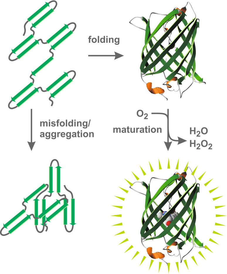
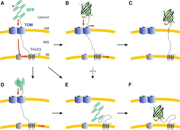
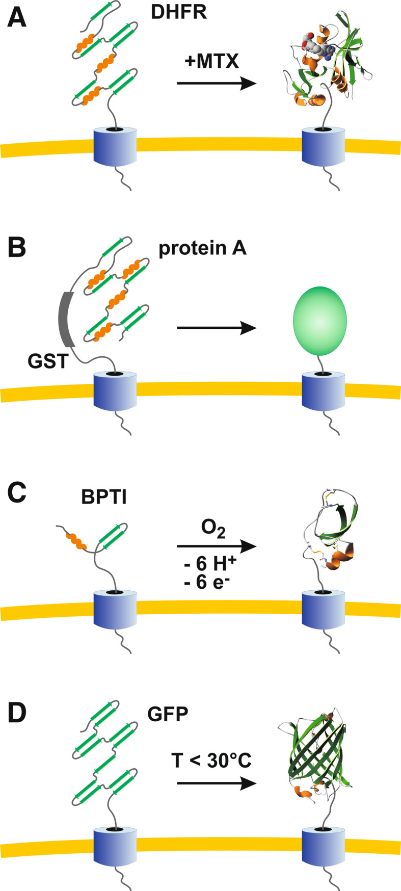
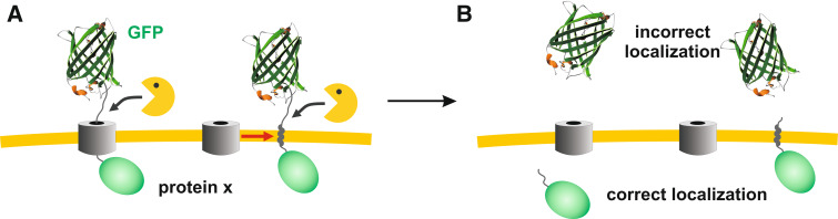
Green fluorescent protein (GFP) is a powerful tool for studying gene expression, protein localization, protein-protein interactions, calcium concentrations, and redox potentials owing to its intrinsic fluorescence. However, GFP not only contains a chromophore but is also tightly folded in a temperature-dependent manner. The latter property of GFP has recently been exploited (1) to characterize the translocase of the outer mitochondrial membrane and (2) to discriminate between protein transport across and into biomembranes in vivo. I therefore suggest that GFP could be a valuable tool for the general analysis of protein transport machineries and pathways in a variety of organisms. Moreover, results from such studies could be important for the interpretation and optimization of classical experiments using GFP tagging.


Figures

Fig. 1
Fig. 1
Properties of GFP. Newly synthesized GFP establishes a stable, near-native tertiary structure at a lower temperature. The protein subsequently matures in the presence of oxygen, yielding the fluorescent chromophore in the center of the beta-barrel. Alternatively, at a higher temperature, GFP tends to misfold and/or to form non-fluorescent aggregates
Fig. 2
Fig. 2
Overview of selected protein transport machineries and their subcellular or suborganellar localizations. TOM, translocase of the outer mitochondrial membrane; TOB/SAM, topogenesis of mitochondrial outer membrane beta-barrel/sorting and assembly machinery; TIM23 and TIM22, translocases of the inner mitochondrial membrane; Oxa1, oxidase assembly insertase; TOC and TIC, translocases of the outer and inner chloroplast envelope; Toc75-V, further member of the Omp85 family; Tat, twin-arginine translocation system; Sec, Sec (secretory) translocase complex; Alb3, albino3 translocase; BAM, beta-barrel assembly machinery; YidC, membrane protein insertase; ERAD, endoplasmic reticulum-associated protein degradation machinery. OM, outer membrane; IMS, intermembrane space; IM, inner membrane; TM, thylakoid membrane; TL, thylakoid lumen; PP, periplasmic space; CP, cytoplasm; EM, ER membrane; PM, peroxisomal membrane
Fig. 3
Fig. 3
Model of the temperature-dependent topology of GFP-Tim23. a After protein synthesis in the cytosol, the C-terminal transmembrane segments of GFP-Tim23 are inserted into the IM via the TOM/TIM22 pathway. b At a lower temperature (e.g., 25°C), GFP folding is faster than protein import. Thus, depending on the hydrophobicity of the OM-spanning segment, threaded GFP-Tim23 either clogs the TOM pore (b) or is laterally inserted into the membrane (c). d At a higher temperature (e.g., 37°C), GFP does not rapidly adopt a stable tertiary structure. Partially folded or aggregated proteins are pulled into the IMS. e Thus, at a higher temperature, the ratios of the GFP-folding-/aggregation-/import-kinetics favor the import of GFP into the IMS. Noteworthy, at a lower temperature, unfolding of the GFP moiety and subsequent import does not seem to occur (as indicated by the dotted arrow), which is in accordance with the stability of GFP in vitro [1, 12, 13]. f Once located in the IMS, GFP-fusion constructs have enough time to adopt a stable conformation
Fig. 4
Fig. 4
Lateral release as a putative physiological rescue mechanism. a Proteins reach the TOM complex in a partially folded state. b Upon protein translocation some proteins might adopt a stable fold before completion of transport. c A lateral release of (random) hydrophobic peptide segments could immediately rescue such clogged TOM pores. Otherwise, the pore would become non-functional (which might result in the necessity to replace the translocase). d A subsequent quality control of outer membrane proteins coupled to proteolytic degradation could remove the mistargeted proteins. One hypothetical advantage of such a mechanism over a direct proteolytic degradation of proteins that remain stuck in the TOM pore is that other (slowly) imported proteins are not accidentally cleaved
Fig. 5
Fig. 5
Comparison of selected protein tags for the analysis of protein transport machineries by blocking protein translocation. a DHFR tagging is highly flexible owing to reversible, MTX-induced stabilization of the protein [41, 42]. b Protein A-tagging (e.g., in combination with GST tagging) is less flexible but allows efficient purification owing to affinity chromatography [46, 47]. c BPTI tagging requires the formation of disulfide bonds and is therefore restricted to oxidizing compartments or in vitro experiments [48]. d GFP tagging (e.g., in combination with His tagging) is flexible owing to temperature-dependent folding kinetics and does not require expensive ligands
Fig. 6
Fig. 6
Potential pitfall using GFP tagging. a Premature folding of GFP-tagged proteins before complete protein translocation might result in membrane-spanning constructs in analogy to GFP-Tim23 (Fig. 3). Such constructs could then be susceptible to endogenous proteases. b The tightly folded GFP moiety, which is highly protease-resistant, subsequently ends up in the wrong compartment, leading, for example, to a cytosolic GFP signal of a mitochondrial protein

Similar articles

Cited by

References

    1. Tsien RY. The green fluorescent protein. Annu Rev Biochem. 1998;67:509–544. doi: 10.1146/annurev.biochem.67.1.509. - DOI - PubMed
    1. Zimmer M. Green fluorescent protein (GFP): applications, structure, and related photophysical behavior. Chem Rev. 2002;102(3):759–781. doi: 10.1021/cr010142r. - DOI - PubMed
    1. Giepmans BN, Adams SR, Ellisman MH, Tsien RY. The fluorescent toolbox for assessing protein location and function. Science. 2006;312(5771):217–224. doi: 10.1126/science.1124618. - DOI - PubMed
    1. Llopis J, McCaffery JM, Miyawaki A, Farquhar MG, Tsien RY. Measurement of cytosolic, mitochondrial, and Golgi pH in single living cells with green fluorescent proteins. Proc Natl Acad Sci USA. 1998;95(12):6803–6808. doi: 10.1073/pnas.95.12.6803. - DOI - PMC - PubMed
    1. Matsuyama S, Llopis J, Deveraux QL, Tsien RY, Reed JC. Changes in intramitochondrial and cytosolic pH: early events that modulate caspase activation during apoptosis. Nat Cell Biol. 2000;2(6):318–325. doi: 10.1038/35014006. - DOI - PubMed

Publication types

Substances

LinkOut - more resources