Review
. 2012 Oct;22(5):785-94.
doi: 10.1016/j.jelekin.2012.01.015. Epub 2012 Feb 19.

Spinal manipulative therapy and somatosensory activation

Affiliations
Review

Spinal manipulative therapy and somatosensory activation

J G Pickar et al. J Electromyogr Kinesiol. 2012 Oct.

Abstract

Manually-applied movement and mobilization of body parts as a healing activity has been used for centuries. A relatively high velocity, low amplitude force applied to the vertebral column with therapeutic intent, referred to as spinal manipulative therapy (SMT), is one such activity. It is most commonly used by chiropractors, but other healthcare practitioners including osteopaths and physiotherapists also perform SMT. The mechanisms responsible for the therapeutic effects of SMT remain unclear. Early theories proposed that the nervous system mediates the effects of SMT. The goal of this article is to briefly update our knowledge regarding several physical characteristics of an applied SMT, and review what is known about the signaling characteristics of sensory neurons innervating the vertebral column in response to spinal manipulation. Based upon the experimental literature, we propose that SMT may produce a sustained change in the synaptic efficacy of central neurons by evoking a high frequency, bursting discharge from several types of dynamically-sensitive, mechanosensitive paraspinal primary afferent neurons.


Figures

Figure 1
Figure 1
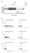
Force time curves derived from high velocity, low amplitude spinal manipulations applied to humans (A & B) and to a simulation device (C). A shows the phases and relative force occurring during the maneuver. (Modified from Fig 1, Herzog W J. Bodywork & Movement Therapies 2010;14:280-6) B shows the mean force-time curves of manipulations applied to the cervical thoracic, and sacroiliac regions. Modified from Fig 9 Herzog W, et al. Spine 1993;18(9):1206-12. C shows the force-time curves of a toggle recoil manipulation performed with a left hand contact (left panel) and a right hand contact (right panel). Modified from Fig 2 Graham BA, et al. Manual Therapy 2010; 15:74-9
Figure 2
Figure 2
Lumbar paraspinal muscle spindle response to a high velocity, low amplitude spinal manipulative-like load. A shows an original tracing of a spindle’s response to the manipulation. Inset shows the spindle’s discharge on an expanded time scale. (Adapted from Journal of Manipulative and Physiological Therapeutics. 24(2):2-11, Pickar J & Wheeler JD, Response of muscle proprioceptors to spinal manipulative-like loads in the anesthetized cat, p6, 2001, with permission from Elsevier) B shows original tracings of a muscle spindle afferent’s response to 6 different thrust durations (400, 200, 100, 50, 25, and 12.5 ms) using a half-sine waveform . (Adapted from Journal of Manipulative and Physiological. Therapeutics. 29(1):22-31, Pickar JG & Kang Y-M, Paraspinal muscle spindle responses to the duration of a spinal manipulation under force control p26, 2006, with permission from Elsevier)
Figure 3
Figure 3
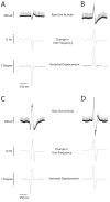
Cervical muscle spindle afferent response to rapid rotation of the C2 vertebra simulating the thrust phase of a high velocity, low amplitude spinal manipulation. In each panel (A, B ,C, D) top trace shows an original recording of spindle activity, middle trace shows the instantaneous discharge frequency (bin width 0.125s) and lower trace shows the C2 vertebra’s relative position. Panel A shows a decrease in the muscle spindle afferent activity during the thrust and panel B shows an increase in its frequency during displacement to the right. The bottom two panels show a muscle spindle’s response to vertebral displacement initially to the left and then right and back to the midline (C) and then with displacement initially to the right and then left and back to the midline (D). Note the DC shift (movement artefact) in the tracing of the original muscle spindle recordings (Raw Unit Activity) is greater when the vertebra is displaced contralaterally to the short dorsal rootlet being recorded. (Bolton PS and Holland CT 1998 unpublished data).

References

    1. Amonoo-Kuofi HS. The density of muscle spindles in the medial, intermediate and lateral columns of human intrinsic postvertebral muscles. Journal of Anatomy. 1983;136(3):509–19. - PMC - PubMed
    1. Bialosky JE, Bishop MD, Price DD, Robinson ME, George SZ. The mechanisms of manual therapy in the treatment of musculoskeletal pain: A comprehensive model. Manual Therapy. 2009;14:531–38. - PMC - PubMed
    1. Bialosky JE, Bishop MD, Robinson ME, Zeppieri G, Jr., George SZ. Spinal manipulative therapy has an immediate effect on thermal pain sensitivity in people with low back pain: a randomized controlled trial. Physical Therapy. 2009;89:1292–1303. - PMC - PubMed
    1. Bishop MD, Beneciuk JM, George SZ. Immediate reduction in temporal sensory summation after thoracic spinal manipulation. The Spine Journal. 2011;11:440–46. - PMC - PubMed
    1. Bolton PS. The somatosensory system of the neck and its effects on the central nervous system. Journal of Manipulative Physiological and Therapeutics. 1998;21(8):553–63. - PubMed

Publication types