Analysis of lumbar pedicle morphology in degenerative spines using multiplanar reconstruction computed tomography: what can be the reliable index for optimal pedicle screw diameter?
- PMID: 22350009
- PMCID: PMC3535235
- DOI: 10.1007/s00586-012-2199-x
Analysis of lumbar pedicle morphology in degenerative spines using multiplanar reconstruction computed tomography: what can be the reliable index for optimal pedicle screw diameter?
Abstract
Purpose: The measurement of transverse pedicle width is still recommended for selecting a screw diameter despite being weakly correlated with the minimum pedicle diameter, except in the upper lumbar spine. The purpose of this study was to reveal the difference between the minimum pedicle diameter and conventional transverse or sagittal pedicle width in degenerative lumbar spines.
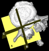
Methods: A total of 50 patients with degenerative lumbar disorders without spondylolysis or lumbar scoliosis of >10° who preoperatively underwent helical CT scans were included. The DICOM data of the scans were reconstructed by imaging software, and the transverse pedicle width (TPW), sagittal pedicle width (SPW), minimum pedicle diameter (MPD), and the cephalocaudal inclination of the pedicles were measured.
Results: The mean TPW/SPW/MPD values were 5.46/11.89/5.09 mm at L1, 5.76/10.44/5.39 mm at L2, 7.25/10.23/6.52 mm at L3, 9.01/9.36/6.83 mm at L4, and 12.86/8.95/7.36 mm at L5. There were significant differences between the TPW and MPD at L3, L4, and L5 (p < 0.01) and between the SPW and MPD at all levels (p < 0.01).
Conclusions: The MPD was significantly smaller than the TPW and SPW at L3, L4, and L5. The actual measurements of the TPW were not appropriate for use as a direct index for the optimal pedicle screw diameter at these levels. Surgeons should be careful in determining pedicle screw diameter based on plain CT scans especially in the lower lumbar spine.
Figures


Similar articles
-
Morphometric analysis using multiplanar reconstructed CT of the lumbar pedicle in patients with degenerative lumbar scoliosis characterized by a Cobb angle of 30° or greater.J Neurosurg Spine. 2012 Sep;17(3):256-62. doi: 10.3171/2012.6.SPINE12227. Epub 2012 Jul 13. J Neurosurg Spine. 2012. PMID: 22794782
-
Pedicle Morphology of Lower Thoracic and Lumbar Spine in Ankylosing Spondylitis Patients with Thoracolumbar Kyphosis: A Comparison with Fracture Patients.Orthop Surg. 2022 Sep;14(9):2188-2194. doi: 10.1111/os.13429. Epub 2022 Aug 16. Orthop Surg. 2022. PMID: 35971839 Free PMC article.
-
Rigid, semirigid versus dynamic instrumentation for degenerative lumbar spinal stenosis: a correlative radiological and clinical analysis of short-term results.Spine (Phila Pa 1976). 2004 Apr 1;29(7):735-42. doi: 10.1097/01.brs.0000112072.83196.0f. Spine (Phila Pa 1976). 2004. PMID: 15087795 Clinical Trial.
-
Congenital lumbar spinal stenosis: a prospective, control-matched, cohort radiographic analysis.Spine J. 2005 Nov-Dec;5(6):615-22. doi: 10.1016/j.spinee.2005.05.385. Spine J. 2005. PMID: 16291100 Clinical Trial.
-
Risk factors for adjacent segment degeneration after PLIF.Spine (Phila Pa 1976). 2004 Jul 15;29(14):1535-40. doi: 10.1097/01.brs.0000131417.93637.9d. Spine (Phila Pa 1976). 2004. PMID: 15247575 Review.
Cited by
-
Influence of Pedicle Screw Insertion Depth on Posterior Lumbar Interbody Fusion: Radiological Significance of Deeper Screw Placement.Global Spine J. 2024 Mar;14(2):470-477. doi: 10.1177/21925682221110142. Epub 2022 Jun 17. Global Spine J. 2024. PMID: 35713986 Free PMC article.
-
Optimal cut-off points of lumbar pedicle thickness as a morphological parameter to predict lumbar spinal stenosis syndrome: a retrospective study.J Pain Res. 2018 Sep 4;11:1709-1714. doi: 10.2147/JPR.S168990. eCollection 2018. J Pain Res. 2018. PMID: 30233228 Free PMC article.
-
Implications of navigation in thoracolumbar pedicle screw placement on screw accuracy and screw diameter/pedicle width ratio.Brain Spine. 2023 Jul 11;3:101780. doi: 10.1016/j.bas.2023.101780. eCollection 2023. Brain Spine. 2023. PMID: 38020982 Free PMC article.
-
Design and radiological confirmation of three-column cortical bone trajectory in the lower thoracic vertebrae.Eur Spine J. 2025 Jun 7. doi: 10.1007/s00586-025-09025-2. Online ahead of print. Eur Spine J. 2025. PMID: 40481841
-
Dynamic Change of Lumbar Structure and Associated Factors: A Retrospective Study.Orthop Surg. 2019 Dec;11(6):1072-1081. doi: 10.1111/os.12557. Epub 2019 Nov 3. Orthop Surg. 2019. PMID: 31679187 Free PMC article.
References
-
- Chadha M, Balain B, Maini L, Dhaon BK. Pedicle morphology of the lower thoracic, lumbar, and S1 vertebrae: an Indian perspective. Spine (Phila Pa 1976) 2003;28:744–749. - PubMed
MeSH terms
LinkOut - more resources
Full Text Sources
Medical

