

A failure of left temporal cortex to specialize for language is an early emerging and fundamental property of autism
- PMID: 22350062
- PMCID: PMC3286331
- DOI: 10.1093/brain/awr364
A failure of left temporal cortex to specialize for language is an early emerging and fundamental property of autism
Abstract
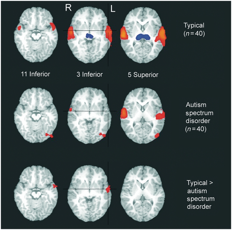
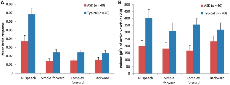
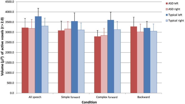
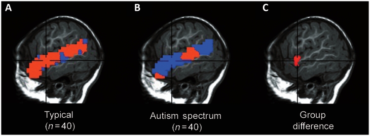
Failure to develop normal language comprehension is an early warning sign of autism, but the neural mechanisms underlying this signature deficit are unknown. This is because of an almost complete absence of functional studies of the autistic brain during early development. Using functional magnetic resonance imaging, we previously observed a trend for abnormally lateralized temporal responses to language (i.e. greater activation on the right, rather than the expected left) in a small sample (n = 12) of sleeping 2-3 year olds with autism in contrast to typically developing children, a finding also reported in autistic adults and adolescents. It was unclear, however, if findings of atypical laterality would be observed in a larger sample, and at even earlier ages in autism, such as around the first birthday. Answers to these questions would provide the foundation for understanding how neurofunctional defects of autism unfold, and provide a foundation for studies using patterns of brain activation as a functional early biomarker of autism. To begin to examine these issues, a prospective, cross-sectional design was used in which brain activity was measured in a large sample of toddlers (n = 80) during the presentation of a bedtime story during natural sleep. Forty toddlers with autism spectrum disorder and 40 typically developing toddlers ranging in age between 12-48 months participated. Any toddler with autism who participated in the imaging experiment prior to final diagnosis was tracked and diagnoses confirmed at a later age. Results indicated that at-risk toddlers later diagnosed as autistic display deficient left hemisphere response to speech sounds and have abnormally right-lateralized temporal cortex response to language; this defect worsens with age, becoming most severe in autistic 3- and 4-year-olds. Typically developing children show opposite developmental trends with a tendency towards greater temporal cortex response with increasing age and maintenance of left-lateralized activation with age. We have now demonstrated lateralized abnormalities of temporal cortex processing of language in autism across two separate samples, including a large sample of young infants who later are diagnosed with autism, suggesting that this pattern may reflect a fundamental early neural developmental pathology in autism.
Figures
Similar articles
-
Neural systems for speech and song in autism.Brain. 2012 Mar;135(Pt 3):961-75. doi: 10.1093/brain/awr335. Epub 2012 Feb 1. Brain. 2012. PMID: 22298195 Free PMC article.
-
Age-related temporal and parietal cortical thinning in autism spectrum disorders.Brain. 2010 Dec;133(Pt 12):3745-54. doi: 10.1093/brain/awq279. Epub 2010 Oct 5. Brain. 2010. PMID: 20926367 Free PMC article.
-
Deviant functional magnetic resonance imaging patterns of brain activity to speech in 2-3-year-old children with autism spectrum disorder.Biol Psychiatry. 2008 Oct 1;64(7):589-98. doi: 10.1016/j.biopsych.2008.05.020. Epub 2008 Jul 30. Biol Psychiatry. 2008. PMID: 18672231 Free PMC article.
-
Update on the language disorders of individuals on the autistic spectrum.Brain Dev. 2003 Apr;25(3):166-72. doi: 10.1016/s0387-7604(02)00191-2. Brain Dev. 2003. PMID: 12689694 Review.
-
Early functional brain development in autism and the promise of sleep fMRI.Brain Res. 2011 Mar 22;1380:162-74. doi: 10.1016/j.brainres.2010.09.028. Epub 2010 Sep 24. Brain Res. 2011. PMID: 20869953 Free PMC article. Review.
Cited by
-
Intrinsic connectivity network mapping in young children during natural sleep.Neuroimage. 2013 Dec;83:288-93. doi: 10.1016/j.neuroimage.2013.05.020. Epub 2013 May 29. Neuroimage. 2013. PMID: 23727317 Free PMC article.
-
Different functional neural substrates for good and poor language outcome in autism.Neuron. 2015 Apr 22;86(2):567-77. doi: 10.1016/j.neuron.2015.03.023. Epub 2015 Apr 9. Neuron. 2015. PMID: 25864635 Free PMC article. Clinical Trial.
-
Atypical hemispheric specialization for faces in infants at risk for autism spectrum disorder.Autism Res. 2015 Apr;8(2):187-98. doi: 10.1002/aur.1438. Epub 2015 Mar 25. Autism Res. 2015. PMID: 25808162 Free PMC article.
-
Age-dependent brain gene expression and copy number anomalies in autism suggest distinct pathological processes at young versus mature ages.PLoS Genet. 2012;8(3):e1002592. doi: 10.1371/journal.pgen.1002592. Epub 2012 Mar 22. PLoS Genet. 2012. PMID: 22457638 Free PMC article.
-
Diffusion Tensor Imaging Provides Evidence of Possible Axonal Overconnectivity in Frontal Lobes in Autism Spectrum Disorder Toddlers.Biol Psychiatry. 2016 Apr 15;79(8):676-84. doi: 10.1016/j.biopsych.2015.06.029. Epub 2015 Jul 4. Biol Psychiatry. 2016. PMID: 26300272 Free PMC article.
References
-
- Anderson AW, Marois R, Colson ER, Peterson BS, Duncan CC, Ehrenkranz RA, et al. Neonatal auditory activation detected by functional magnetic resonance imaging. Magn Reson Imaging. 2001;19:1–5. - PubMed
-
- Anderson B, Southern BD, Powers RE. Anatomic asymmetries of the posterior superior temporal lobes: a postmortem study. Neuropsychiatry Neuropsychol Behav Neurol. 1999;12:247–54. - PubMed
-
- Bartak L, Rutter M, Cox A. A comparative study of infantile autism and specific development receptive language disorder. I. The children. Br J Psychiatry. 1975;126:127–45. - PubMed
