. 2012 Apr 1;72(7):1878-89.
doi: 10.1158/0008-5472.CAN-11-3132. Epub 2012 Feb 20.

Pten loss and RAS/MAPK activation cooperate to promote EMT and metastasis initiated from prostate cancer stem/progenitor cells

Affiliations

Pten loss and RAS/MAPK activation cooperate to promote EMT and metastasis initiated from prostate cancer stem/progenitor cells

David J Mulholland et al. Cancer Res. .

Abstract

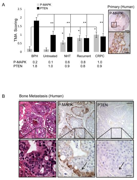
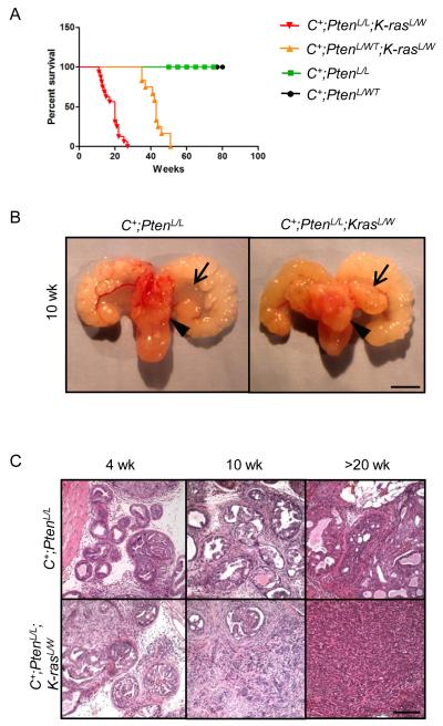
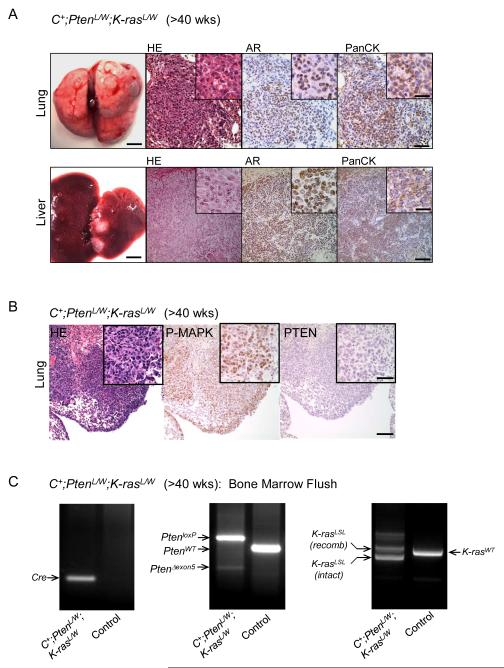
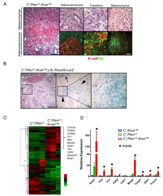
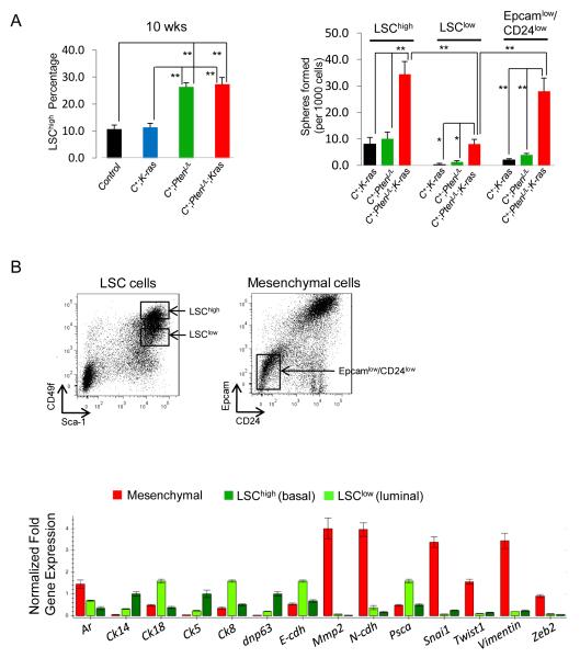
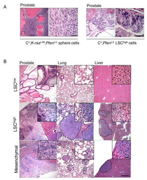
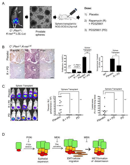
PTEN loss or PI3K/AKT signaling pathway activation correlates with human prostate cancer progression and metastasis. However, in preclinical murine models, deletion of Pten alone fails to mimic the significant metastatic burden that frequently accompanies the end stage of human disease. To identify additional pathway alterations that cooperate with PTEN loss in prostate cancer progression, we surveyed human prostate cancer tissue microarrays and found that the RAS/MAPK pathway is significantly elevated in both primary and metastatic lesions. In an attempt to model this event, we crossed conditional activatable K-ras(G12D/WT) mice with the prostate conditional Pten deletion model. Although RAS activation alone cannot initiate prostate cancer development, it significantly accelerated progression caused by PTEN loss, accompanied by epithelial-to-mesenchymal transition (EMT) and macrometastasis with 100% penetrance. A novel stem/progenitor subpopulation with mesenchymal characteristics was isolated from the compound mutant prostates, which was highly metastatic upon orthotopic transplantation. Importantly, inhibition of RAS/MAPK signaling by PD325901, a mitogen-activated protein (MAP)-extracellular signal-regulated (ER) kinase (MEK) inhibitor, significantly reduced the metastatic progression initiated from transplanted stem/progenitor cells. Collectively, our findings indicate that activation of RAS/MAPK signaling serves as a potentiating second hit to alteration of the PTEN/PI3K/AKT axis, and cotargeting both the pathways is highly effective in preventing the development of metastatic prostate cancers.


Figures

Figure 1
Figure 1. RAS/MAPK signaling is enriched in human prostate cancer
A, PTEN and P-MAPK expression in human TMAs (patient samples = 194, cores = 388) (left) and P-MAPK expression in recurrent Prostate cancer (right), *, p<0.05, low mag bar = 1 mm, high mag bar = 100 μm. B, Expression of PTEN and P-MAPK in human bone metastasis. low mag bar = 250 μm, high mag bar = 50 μm.
Figure 2
Figure 2. Pten loss and Ras activation cooperate to enhance murine prostate cancer progression
A, Kaplan Meier survival curve of Pten and Pten;K-ras mutants. B, Gross structure of intact C+;PtenL/L and C+;PtenL/L;K-rasL/W mutant prostates at 10 wks (arrow head = lateral lobe, arrow = anterior lobe), bar = 4 mm. C, Histology of intact C+;PtenL/L and C+;PtenL/L;K-rasL/W mutant prostates at 4, 10 and >20 wks, bar = 250 μm.
Figure 3
Figure 3. Pten loss and Ras activation cooperate to significantly enhance metastatic burden
A, Gross lung and liver structure showing the presence of macrometastasis and corresponding stains for HE, androgen receptor (AR) and pan-cytokeratin (PanCK) in C+;PtenL/WT;K-rasL/W mutants (>40 wks). Low mag bar = 150 μm, high mag bar = 50 μm. B, Lung lesions from C+;PtenL/W;K-rasL/W mutants showing P-MAPK and PTEN expression. Low mag bar = 150 μm, high mag bar = 50 μm. C, Bone marrow flush and PCR genotyping for excised Pten (Δexon5) and recombined LSL-K-Ras in C+;PtenL/W;K-rasL/WT and control (WT) mice.
Figure 4
Figure 4. Pten loss and Ras pathway activation propagate an EMT signature
A, Histology (left, top) and immunostains (E-cadherin, Vimentin) (bottom) showing regions of transition between epithelial and mesenchymal phenotypes, low mag bar = 500 μm, high mar bar = 100 μm. B, Lineage tracing using beta-gal staining and the LSL-Rosa26-LacZ reporter in conjunction with the epithelial specific probasin promoter in C+;PtenL/L;K-rasL/W mutants (10 wks), low mag bar = 500 μm, high mar bar = 200 μm. C, Gene microarray analysis showing EMT pathway gene activity in between C+;PtenL/L;K-rasL/W and C+;PtenL/L mutants. D, RT-PCR confirmation of EMT gene alterations in C+;PtenL/L;K-rasL/W mutant prostates (*, p < 0.05).
Figure 5
Figure 5. C+;PtenL/L;K-rasL/W mutant LSChigh and mesenchymal cells show high stem/progenitor activity
A, Comparison of percentage LSChigh subpopulation in control, C+;K-rasL/W C+;PtenL/L and C+;PtenL/L;K-rasL/W mutant prostates (10 wks) (Left panel). Comparison of sphere plating efficiency between LSChigh, LSClow and mesenchymal cells isolated from C+;PtenL/L and C+;PtenL/L;K-rasL/W mutant prostates (10 wks) (*, p < 0.05; **, p<0.01) (Right panel). B, Isolation of LSChigh(LinSca1+CD49fhigh), LSClow (LinSca1+CD49flow) and mesenchymal cells from C+;PtenL/L;K-rasL/W mutants (10 wks) with RT-PCR analysis (lower panel)
Figure 6
Figure 6. Transplantation of C+;PtenL/L;K-rasL/W stem/progenitor cells are sufficient to initiate EMT and metastasis
A, Orthotopic transplantation of C+;PtenL/L;K-rasL/W spheres cells (left panel) and C+;PtenL/L LSChigh cells (right panel) to NOD;SCID;IL2rγ-null recipients and resulting pathology (left). B, Transplantation of LSClow, LSChigh and mesenchymal cells isolated from primary C+;PtenL/L;K-rasL/W mutant cells and resulting pathology observed in NOD;SCID;IL2rγ-null recipients. Low mag bar = 500 μm, high mag bar = 100 μm.
Figure 7
Figure 7. Pharmacological targeting of RAS/MAPK signaling inhibits metastatic disease initiated from C+;PtenL/L;K-rasL/W mutant stem/progenitor cells
A, Isolation of prostate sphere cells from C+;PtenL/L;K-rasL/W;LSL-Luc mutants and orthotopic injection to NOD;SCID;IL2rγ-null mice. Recipients were then treated with either placebo, rapamycin (R) + PD325901 (PD) or PD325901, alone. B, Effect of rapamycin/PD325901 treatment on P-MAPK and P-S6 levels (left panel), cell proliferation (Ki67+ index; middle panel) and mesenchymal content (right panel). C, Effect of rapamycin/PD325901 or PD325901 on thoracic region BLI (left, middle panels) and metastatic lung lesion content (right panel). D, Model showing that Pten-null LSChigh cells can initiate prostate cancer and with RAS/MAPK activation lead to EMT, metastatic disease and formation of macrometastatic lesions.

References

    1. ACS American Cancer Society. Cancer Facts & Figures. 2010
    1. Taylor BS, Schultz N, Hieronymus H, et al. Integrative genomic profiling of human prostate cancer. Cancer Cell. 2010;18:11–22. - PMC - PubMed
    1. Wang S, Gao J, Lei Q, et al. Prostate-specific deletion of the murine Pten tumor suppressor gene leads to metastatic prostate cancer. Cancer Cell. 2003;4:209–21. - PubMed
    1. Svensson RU, Haverkamp JM, Thedens DR, Cohen MB, Ratliff TL, Henry MD. Slow disease progression in a C57BL/6 pten-deficient mouse model of prostate cancer. Am J Pathol. 2011;179:502–12. - PMC - PubMed
    1. Ma X, Ziel-van der Made AC, Autar B, et al. Targeted biallelic inactivation of Pten in the mouse prostate leads to prostate cancer accompanied by increased epithelial cell proliferation but not by reduced apoptosis. Cancer Res. 2005;65:5730–9. - PubMed

Publication types

MeSH terms