

doi: 10.1007/s10858-012-9613-x.
Epub 2012 Feb 17.
High dimensional and high resolution pulse sequences for backbone resonance assignment of intrinsically disordered proteins
Affiliations
- PMID: 22350953
- PMCID: PMC3315646
- DOI: 10.1007/s10858-012-9613-x
Item in Clipboard
High dimensional and high resolution pulse sequences for backbone resonance assignment of intrinsically disordered proteins
J Biomol NMR.
2012 Apr.
Abstract
Four novel 5D (HACA(N)CONH, HNCOCACB, (HACA)CON(CA)CONH, (H)NCO(NCA)CONH), and one 6D ((H)NCO(N)CACONH) NMR pulse sequences are proposed. The new experiments employ non-uniform sampling that enables achieving high resolution in indirectly detected dimensions. The experiments facilitate resonance assignment of intrinsically disordered proteins. The novel pulse sequences were successfully tested using δ subunit (20 kDa) of Bacillus subtilis RNA polymerase that has an 81-amino acid disordered part containing various repetitive sequences.
Figures

Correlation of HA–CA (a), HB–CB (b), and N–CO (c) chemical shifts for the 81 a.a. unstructured part of the δ subunit of B. subtilis RNA polymerase. The 1H and 13C chemical shifts depend mostly on particular amino acid residue, whereas 13CO and 15N frequencies are much better resolved and enable identification of backbone connectivity. (BMRB entry 16912)
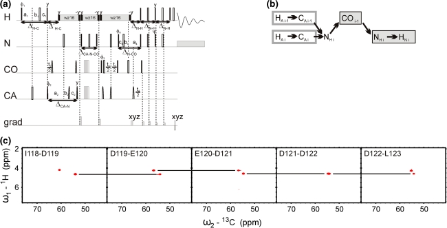
5D HACA(N)CONH technique. (a) Pulse sequence, 1H, 13CA, and 15N evolution is in semi-constant-time mode: ai = (t
i + Δ)/2, b
i = t
i(1−Δ/t
imax)/2, c
i = Δ(1−t
i/t
imax)/2 (where Δ stands for coherence transfer delays listed below, t
i is the evolution time in ith dimension, and t
imax is maximal length of the evolution time delay). Delays were set as follows: Δ’H-C = 2.6 ms, ΔCA-N = 28.0 ms, ΔCA-N-CO = 28.0 ms, ΔN-CO = 28.0 ms, and ΔN-H = 5.4 ms. The four-step phase cycle was used: ϕ1 = x, −x, ϕ2 = 2x, 2(−x) and Rec = ϕ1 + ϕ2. Simultaneous inversion of CA and CO spins was achieved using 6-element composite pulse (Shaka 1985). The coherence selection gradients (marked xyz) were applied at the magic angle. The phase ψ was inverted simultaneously with the last gradient pulse. (b) Coherence transfer in the peptide chain. HN, N, and CO frequencies (filled rectangles) are ‘fixed’ for Fourier transform. Frames for HA and CA indicate the dimensions of 2D cross-sections obtained by SMFT procedure. (c) 2D spectral planes for the δ subunit of B. subtilis RNA polymerase, which were obtained by SMFT procedure performed on the 5D randomly sampled signal (Poisson disk sampling) with ‘fixed’ frequencies obtained from 3D HNCO peak list. Each cross-section contains two cross-peaks: for’fixed’ HiN, Ni and COi−1, the peaks correspond to HAi–CAi and HAi−1–CAi−1 correlations
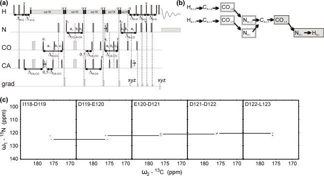
5D (HACA)CON(CA)CONH technique. (a) Pulse sequence, 15N (in t
2 and t
4) and 13CO (in t
1 and t
3) evolution is in semi-constant-time mode: ai = (t
i + Δ)/2, b
i = t
i(1−Δ/t
imax)/2, c
i = Δ(1−t
i/t
imax)/2 (where Δ stands for coherence transfer delays listed below, t
i is the evolution time in ith dimension, and t
imax is maximal length of the evolution time delay). Delays were set as follows: ΔH-C = 3.7 ms, Δ’H-C = 2.6 ms, ΔCA-CO = 6.8 ms, ΔCA-N = 28.0 ms, ΔCA-N-CO = 28.0 ms, ΔN-CO = 28.0 ms, ΔN-H = 5.4 ms. The four-step phase cycle was used: ϕ1 = x, −x, ϕ2 = 2x, 2(−x) and Rec = ϕ1 + ϕ2. Simultaneous inversion of CA and CO spins was achieved using 6-element composite pulse (Shaka 1985). The coherence selection gradients (marked by xyz) were applied at the magic angle. The phase ψ was inverted simultaneously with the last gradient pulse. (b) Coherence transfer in the peptide chain. HN, N, and CO frequencies (filled rectangles) are ‘fixed’ for Fourier transform. Frames for N and CO indicate the dimensions of 2D cross-sections obtained by SMFT procedure. (c) 2D spectral planes for the δ subunit of B. subtilis RNA polymerase, which were obtained by SMFT procedure performed on the 5D randomly sampled signal (Poisson disk sampling) with ‘fixed’ frequencies obtained from 3D HNCO peak list. Each cross-section contains two cross-peaks: for ‘fixed’ HiN, Ni and COi−1 the peaks correspond to Ni-COi−1 and Ni−1-COi−2 correlations
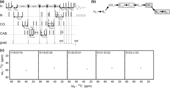
5D HNCOCACB technique. (a) Pulse sequence, 13CO and 15N evolution is in semi-constant-time mode: ai = (t
i + Δ)/2, b
i = t
i(1−Δ/t
imax)/2, c
i = Δ(1−t
i/t
imax)/2 (where Δ stands for listed below coherence transfer delays, t
i is the evolution time in ith dimension and t
imax is maximal length of the evolution time delay). 13CA chemical shift evolution is in constant-time mode. Delays were set as follows: ΔN-H = 5.4 ms, ΔN-CO = 28.0 ms, ΔCA-CO = 9.1 ms, and ΔCACB = 14.3 ms. The four-step phase cycle was used: ϕ1 = x, −x, ϕ2 = 2x, 2(−x) and Rec = ϕ1 + ϕ2. The coherence selection gradients (marked by xyz) were applied at the magic angle. The phase ψ was inverted simultaneously with the last gradient pulse. (b) Coherence transfer in the peptide chain. HN, N, and CO frequencies (filled rectangles) are ‘fixed’ for Fourier transform. Frames for CA and CB indicate the dimensions of 2D cross-sections obtained by SMFT procedure. (c) 2D spectral planes for the δ subunit of B. subtilis RNA polymerase, which were obtained by SMFT procedure performed on the 5D randomly sampled signal (Poisson disk sampling) with ‘fixed’ frequencies obtained from 3D HNCO peak list. Each cross-section contains one cross-peak: for ‘fixed’ HiN, Ni and COi−1 the peak corresponds to CAi−1-CBi−1 correlation
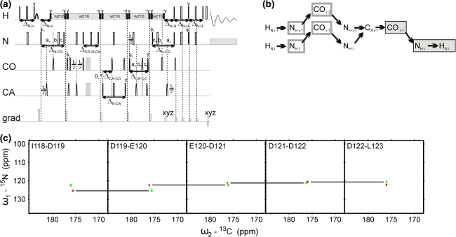
5D (H)NCO(NCA)CONH technique. (a) Pulse sequence, 15N (in t
1 and t
4) and 13CO (in t
3) evolution is in semi-constant-time mode: ai = (t
i + Δ)/2, b
i = t
i(1−Δ/t
imax)/2, c
i = Δ(1−t
i/t
imax)/2 (where Δ stands for listed below coherence transfer delays, t
i is the evolution time in ith dimension and t
imax is maximal length of the evolution time delay). Delays were set as follows: ΔN-H = 5.4 ms, ΔN-CO = 28.0 ms, ΔCO–N-CA = 28.0 ms, ΔN-CA = 28.6 ms, and ΔCA-CO = 9.1 ms. The four-step phase cycle was used: ϕ1 = x, −x, ϕ2 = 2x, 2(−x) and Rec = ϕ1 + ϕ2. Simultaneous inversion of CA and CO spins was achieved using 6-element composite pulse (Shaka 1985). The coherence selection gradients (marked by xyz) were applied at the magic angle. The phase ψ was inverted simultaneously with the last gradient pulse. (b) Coherence transfer in the peptide chain. HN, N, and CO frequencies (filled rectangles) are ‘fixed’ for Fourier transform. Frames for N and CO indicate the dimensions of 2D cross-sections obtained by SMFT procedure. (c) 2D spectral planes for the δ subunit of B. subtilis RNA polymerase, which were obtained by SMFT procedure performed on the 5D randomly sampled signal (Poisson disk sampling) with ‘fixed’ frequencies obtained from 3D HNCO peak list. Each cross-section contains two cross-peaks: for ‘fixed’ HiN, Ni and COi−1 the peaks correspond to Ni-COi−1 and Ni−1-COi−2 correlations

6D (H)NCO(N)CACONH technique. (a) Pulse sequence, 15N (in t
1 and t
5) and 13CO (in t
4) evolution is in semi-constant-time mode: ai = (t
i + Δ)/2, b
i = t
i(1−Δ/t
imax)/2, c
i = Δ(1−t
i/t
imax)/2 (where Δ stands for listed below coherence transfer delays, t
i is the evolution time in ith dimension and t
imax is maximal length of the evolution time delay). CA evolution (in t
3) is in constant-time mode. Delays were set as follows: ΔN-H = 5.4 ms, ΔN-CO = 28.0 ms, ΔCO-N-CA = 28.0 ms, ΔN-CA = 28.6 ms, and ΔCA-CO = 9.1 ms. The four-step phase cycle was used: ϕ1 = x, −x, ϕ2 = 2x, 2(−x) and Rec = ϕ1 + ϕ2. Simultaneous inversion of CA and CO spins was achieved using 6-element composite pulse (Shaka 1985). The coherence selection gradients (marked by xyz) were applied at the magic angle. The phase ψ was inverted simultaneously with the last gradient pulse. (b) Coherence transfer in the peptide chain. HN, N, CO and CA frequencies (filled rectangles) are ‘fixed’ for Fourier transform. Frames for N and CO indicate the dimensions of 2D cross-sections obtained by SMFT procedure. (c) 2D spectral planes for the δ subunit of B. subtilis RNA polymerase, which were obtained by SMFT procedure performed on the 6D randomly sampled signal (Poisson disk sampling) with ‘fixed’ frequencies obtained from 4D HNCOCA peak list. Each plane contains two cross-peaks: for ‘fixed’ HiN, Ni, COi−1 and CAi−1 the peaks correspond to Ni-COi−1 and Ni−1-COi−2 correlations
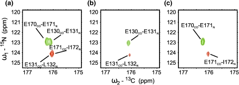
Comparison of 2D N-CO cross-sections from 5D (H)NCO(NCA)CONH (a) and 6D (H)NCO(N)CACONH (b, c) experiments. In 5D experiment (a) the two pairs of correlation peaks overlap (E130/E131 with E170/E171, and E131/L132 with E171/I172) due to similarity of chemical shifts for all ‘fixed’ dimensions: HL132N and HI172N, NL132 and NI172, COE131 and COE171. The additional CA dimension in 6D experiment (b, c) enabled to differentiate and assign all peaks
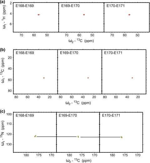
F
1–F
2 2D cross-sections of HACA(N)CONH (a), HNCOCACB (b), and (H)NCO(NCA)CONH (c) 5D experiments, acquired for the δ subunit of B. subtilis RNA polymerase, show E168-E171correlations. The two intra- and inter-residual HA-CA correlation peaks are not resolved in (a); CA-CB correlation peaks in (b) have identical coordinates, however, the resolved pairs of N-CO peaks shown in (c) enable unambiguous sequential assignment
References
-
- Bermel W, Bertini I, Felli IC, Piccioli M, Pierattelli R. 13C detected protonless NMR spectroscopy of proteins in solution. Prog Nucl Magn Reson Spectrosc. 2006;48:25–45. doi: 10.1016/j.pnmrs.2005.09.002. - DOI
Publication types
MeSH terms
Substances
LinkOut - more resources
Full Text Sources
Molecular Biology Databases
