

Peripheral vasodilatation determines cardiac output in exercising humans: insight from atrial pacing
- PMID: 22351638
- PMCID: PMC3573320
- DOI: 10.1113/jphysiol.2011.225334
Peripheral vasodilatation determines cardiac output in exercising humans: insight from atrial pacing
Abstract
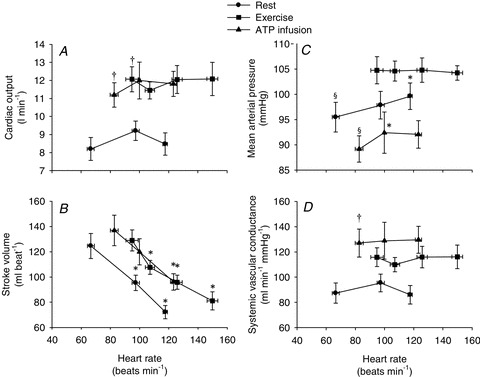
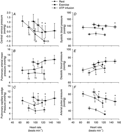
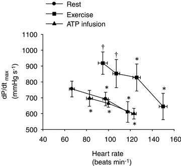
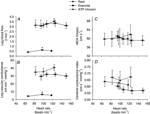
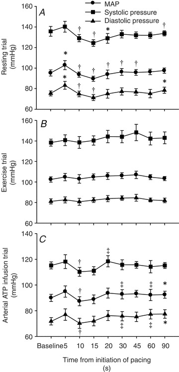
In dogs, manipulation of heart rate has no effect on the exercise-induced increase in cardiac output. Whether these findings apply to humans remain uncertain, because of the large differences in cardiovascular anatomy and regulation. To investigate the role of heart rate and peripheral vasodilatation in the regulation of cardiac output during steady-state exercise, we measured central and peripheral haemodynamics in 10 healthy male subjects, with and without atrial pacing (100–150 beats min(−1)) during: (i) resting conditions, (ii) one-legged knee extensor exercise (24 W) and (iii) femoral arterial ATP infusion at rest. Exercise and ATP infusion increased cardiac output, leg blood flow and vascular conductance (P < 0.05), whereas cerebral perfusion remained unchanged. During atrial pacing increasing heart rate by up to 54 beats min(−1), cardiac output did not change in any of the three conditions, because of a parallel decrease in stroke volume (P < 0.01). Atrial pacing increased mean arterial pressure (MAP) at rest and during ATP infusion (P < 0.05), whereas MAP remained unchanged during exercise. Atrial pacing lowered central venous pressure (P < 0.05) and pulmonary capillary wedge pressure (P < 0.05) in all conditions, whereas it did not affect pulmonary mean arterial pressure. Atrial pacing lowered the left ventricular contractility index (dP/dt) (P < 0.05) in all conditions and plasma noradrenaline levels at rest (P < 0.05), but not during exercise and ATP infusion. These results demonstrate that the elevated cardiac output during steady-state exercise is regulated by the increase in skeletal muscle blood flow and venous return to the heart, whereas the increase in heart rate appears to be secondary to the regulation of cardiac output.
Figures
Comment in
-
Exercise: where the body leads and the heart must follow.J Physiol. 2012 Sep 1;590(17):4127-8. doi: 10.1113/jphysiol.2012.239285. J Physiol. 2012. PMID: 22962034 Free PMC article. No abstract available.
-
Central versus peripheral control of cardiac output in humans: insight from atrial pacing.J Physiol. 2012 Oct 15;590(20):4977-8. doi: 10.1113/jphysiol.2012.240143. J Physiol. 2012. PMID: 23082024 Free PMC article. No abstract available.
Similar articles
-
Skeletal muscle signaling and the heart rate and blood pressure response to exercise: insight from heart rate pacing during exercise with a trained and a deconditioned muscle group.Hypertension. 2013 May;61(5):1126-33. doi: 10.1161/HYPERTENSIONAHA.111.00328. Epub 2013 Mar 11. Hypertension. 2013. PMID: 23478101
-
Haemodynamic responses to exercise, ATP infusion and thigh compression in humans: insight into the role of muscle mechanisms on cardiovascular function.J Physiol. 2008 May 1;586(9):2405-17. doi: 10.1113/jphysiol.2008.152058. Epub 2008 Mar 13. J Physiol. 2008. PMID: 18339690 Free PMC article.
-
On the contribution of group III and IV muscle afferents to the circulatory response to rhythmic exercise in humans.J Physiol. 2011 Aug 1;589(Pt 15):3855-66. doi: 10.1113/jphysiol.2011.209353. Epub 2011 Jun 6. J Physiol. 2011. PMID: 21646407 Free PMC article.
-
[Value of training-induced effects on arterial vascular system and skeletal muscles in therapy of NYHA II/III heart failure].Z Kardiol. 2001 Nov;90(11):813-23. doi: 10.1007/s003920170080. Z Kardiol. 2001. PMID: 11771449 Review. German.
-
A critical assessment of sympathetic restraint in submaximal exercise: Implications for integrated cardiovascular circuit control in exercise.Exp Physiol. 2025 May;110(5):708-721. doi: 10.1113/EP091436. Epub 2025 Mar 25. Exp Physiol. 2025. PMID: 40131015 Free PMC article. Review.
Cited by
-
Possible Effects of Beetroot Supplementation on Physical Performance Through Metabolic, Neuroendocrine, and Antioxidant Mechanisms: A Narrative Review of the Literature.Front Nutr. 2021 May 13;8:660150. doi: 10.3389/fnut.2021.660150. eCollection 2021. Front Nutr. 2021. PMID: 34055855 Free PMC article. Review.
-
Effect of increasing heart rate on finger photoplethysmography fitness index (PPGF) in subjects with implanted cardiac pacemakers.PLoS One. 2018 Nov 28;13(11):e0207301. doi: 10.1371/journal.pone.0207301. eCollection 2018. PLoS One. 2018. PMID: 30485318 Free PMC article. Clinical Trial.
-
Dehydration reduces stroke volume and cardiac output during exercise because of impaired cardiac filling and venous return, not left ventricular function.Physiol Rep. 2020 Jun;8(11):e14433. doi: 10.14814/phy2.14433. Physiol Rep. 2020. PMID: 32538549 Free PMC article.
-
The heart, a secondary organ in the control of blood circulation.Exp Physiol. 2025 May;110(5):649-665. doi: 10.1113/EP091387. Epub 2023 Dec 21. Exp Physiol. 2025. PMID: 38126953 Free PMC article. Review.
-
The cardiovascular changes underlying a low cardiac output with exercise in patients with type 2 diabetes mellitus.Front Physiol. 2024 Mar 20;15:1294369. doi: 10.3389/fphys.2024.1294369. eCollection 2024. Front Physiol. 2024. PMID: 38571722 Free PMC article. Review.
References
-
- Åstrand PO, Cuddy TE, Saltin B, Stenberg J. Cardiac output during submaxial and maximal work. J Appl Physiol. 1964;19:268–274. - PubMed
-
- Bevegård S, Jonsson B, Karlof I, Lagergren H, Sowton E. Effect of changes in ventricular rate on cardiac output and central pressures at rest and during exercise in patients with artificial pacemakers. Cardiovasc Res. 1967;1:21–33. - PubMed
-
- Boushel R, Calbet JA, Rådegran G, Sondergaard H, Wagner PD, Saltin B. Parasympathetic neural activity accounts for the lowering of exercise heart rate at high altitude. Circulation. 2001;104:1785–1791. - PubMed
Publication types
MeSH terms
Substances
LinkOut - more resources
Full Text Sources
Medical
Miscellaneous