Review
. 2012 Mar;25(3):427-42.
doi: 10.1002/nbm.1633. Epub 2011 Feb 20.

New advances in MR-compatible bioartificial liver

Affiliations
Review

New advances in MR-compatible bioartificial liver

Rex E Jeffries et al. NMR Biomed. 2012 Mar.

Abstract

MR-compatible bioartificial liver (BAL) studies have been performed for 30 years and are reviewed. There are two types of study: (i) metabolism and drug studies using multinuclear MRS; primarily short-term (< 8 h) studies; (ii) the use of multinuclear MRS and MRI to noninvasively define the features and functions of BAL systems for long-term liver tissue engineering. In the latter, these systems often undergo not only modification of the perfusion system, but also the construction of MR radiofrequency probes around the bioreactor. We present novel MR-compatible BALs and the use of multinuclear MRS ((13)C, (19)F, (31)P) for the noninvasive monitoring of their growth, metabolism and viability, as well as (1)H MRI methods for the determination of flow profiles, diffusion, cell distribution, quality assurance and bioreactor integrity. Finally, a simple flexible coil design and circuit, and life support system, are described that can make almost any BAL MR-compatible.


Figures

Figure 1
Figure 1
The three bioartificial liver (BAL) designs described: hybrid hollow fiber–microcarrier bioreactor; multicoaxial hollow fiber membrane bioreactor; and fluidized-bed encapsulation bioreactor. Three photographs of the bioreactors (A) with details of their design (B) and flow patterns (C). ECC, extracapillary compartment; ICC, intracapillary compartment.
Figure 2
Figure 2
Generic bioreactor life-support loop and bioreactor. Block diagram of the bioreactor loop showing the major components and the gas exchange module that heats and oxygenates the medium simultaneously (12), so that it can be maintained at room temperature.
Figure 3
Figure 3
MR compatibilization of bioartificial livers (BALs), showing the insertion of the assembly into the bottom of a wide-bore 400-MHz NMR spectrometer (A) and the photo-etched flexible Helmholtz copper coils (B) that are wrapped around the hybrid hollow fiber–microcarrier BAL to serve as the low-frequency coil, increasing the filling factor; this is then inserted into the outer high band coil wrapped on two semicircular glass cylinders and soldered to the circuit in the background (C). The BAL–loop–MR probe–cradle assembly is inserted into an aluminum radiofrequency (RF) shield (D and E), or a larger imaging probe, either commercial or home-built, can be used to obtain images, as shown by the 1H/23Na NMR probe (F).
Figure 4
Figure 4
(A) Inoculation of the hybrid hollow fiber bioreactor with hepatocytes mixed with microcarriers. (B) Graphs of the percentage change in β-nucleotide triphosphate (β-NTP) relative to β-NTP from the first spectrum of hepatocytes perfused with medium subjected to 20% and 40% oxygen, and 20% oxygen but directly perfused through the cell compartment. (C) Analysis of the medium components after 20% and 40% oxygen treatments with a regular flow configuration, showing the increase in lactate dehydrogenase (LDH), validating that the decrease in β-NTP is a result of cell death (glucose and ammonia are also quantified).
Figure 5
Figure 5
19F MR spectral time course of the metabolic probe, 2-fluoroacetanilide, and its metabolite, 2-fluoroaniline, with their structures and metabolic pathway (top). Each spectrum required 1 h to acquire. The hybrid hollow fiber was used and inoculated with microcarriers coated with rat hepatocytes and perfused with Dulbecco's modified Eagle's medium oxygenated with 20% oxygen : 5% carbon dioxide : 75% nitrogen. The fluorinated metabolic probe was added to the medium after 24 h of 31P acquisition, as shown in Fig. 4B for 20% oxygen treatment (top graph); therefore, the time course starts at 25 h post-inoculation.
Figure 6
Figure 6
(A) Transaxial T2-weighted MRI of two coaxial hollow fiber prototypes, with 18 (left) and 40 (right) fiber pairs with diffusion distances of 500 and 200 μm, respectively. (B) The enlargement of a partially (left) and completely (right) filled fiber pair from the 18- and 40-fiber pair prototypes. The effect of flow results in hypointensity in the intracapillary compartment (ICC) (B, right image). Modifications of images from Macdonald et al. (29).
Figure 7
Figure 7
(A) Transaxial velocity-encoded MRI of the multicoaxial hollow fiber bioreactor (inset) at two inward radial flow rates. Note the difference in flow of the extracapillary compartment (ECC) between the two flow rates, and that the intracapillary compartment (ICC) flow is out of the range of velocities calibrated for the MRI experiment and is thus folded. The FIDAP (Fluid Dynamics Analysis Package) theoretical analysis of flow in the three compartments is given in (B). Note that the laminar nature matches the ECC and cell compartments, and predicts a higher velocity profile in the ECC. Qrad, radial flow. Reproduced by permission from Wolfe et al. (33).
Figure 8
Figure 8
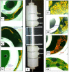
Confocal cross-sectional images of the coaxial bioreactor at culture day 30. (A1–A3) Cells embedded in collagen : Matrigel. (B) Generation-1 coaxial bioreactor. (C1–C4) Cells cultured with alginate : Matrigel beads. Note that the cells are aggregated in A1–A3, but not as extensively in C2–C4, except along the borders of the alginate beads (C1).
Figure 9
Figure 9
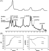
(A) Comparison of in vivo 31P MR spectra from the hollow fiber membrane bioreactor (29) and fluidized-bed bioreactor (74) obtained in 60 min vs 30 min, demonstrating the superior signal-to-noise ratio (SNR) and resolution of the fluidized-bed bioreactor. (B) Graph of intracellular pH (left) and β-nucleotide triphosphate (β-NTP) (right) of rat hepatocytes perfused with medium subjected to 20% and 35% oxygen treatments. GPC, glycerophosphocholine; NAD(H), nicotinamide adenine dinucleotide; Pi, inorganic phosphate; PME, phosphomonoester; UDPG, uridine diphosphoglucose.
Figure 10
Figure 10
(A) The 18-h in vivo 13C MR spectral time course of the fluidized-bed bioreactor inoculated with 500-μm-diameter electrostatically encapsulated alginate beads containing 3.5 × 10⁷ rat hepatocytes/mL and perfused with 25 mm u-13C-glucose, showing the increase in peak representing the C1-glycogen at 102 ppm. (B) in vivo 13C MR spectrum of perfused alginate-encapsulated JM-1 cells and the 15-h time course of C2-lactate above the spectrum.

Similar articles

Cited by

References

    1. Dale BE, Gillies RJ. Nuclear magnetic resonance spectroscopy of dense cell populations for metabolic studies and bioreactor engineering: a synergistic partnership. Biotechnology. 1991;17:107–118. - PubMed
    1. Ruiz-Cabello J, Cohen JS. NMR and the study of pathological state in cells and tissues. Int. Rev. Cytol. 1993;145:1–63. - PubMed
    1. Kaplan O, Cohen JS. Metabolism of breast cancer cells as revealed by non-invasive magnetic resonance spectroscopy studies. Breast Cancer Res. Treat. 1994;31(2–3):285–299. - PubMed
    1. Szwergold BS. NMR spectroscopy of cells. Annu. Rev. Physiol. 1992;54:775–798. - PubMed
    1. Zupke C, Foy B. Nuclear magnetic resonance analysis of cell metabolism. Curr. Opin. Biotechnol. 1995;6(2):192–197. - PubMed

Publication types