

Tumor epidermal growth factor receptor and EGFR PY1068 are independent prognostic indicators for head and neck squamous cell carcinoma
- PMID: 22351687
- PMCID: PMC3430124
- DOI: 10.1158/1078-0432.CCR-11-1593
Tumor epidermal growth factor receptor and EGFR PY1068 are independent prognostic indicators for head and neck squamous cell carcinoma
Abstract
Purpose: To assess the prognostic value of epidermal growth factor receptor (EGFR) molecular characteristics of head and neck squamous cell carcinoma (HNSCC).
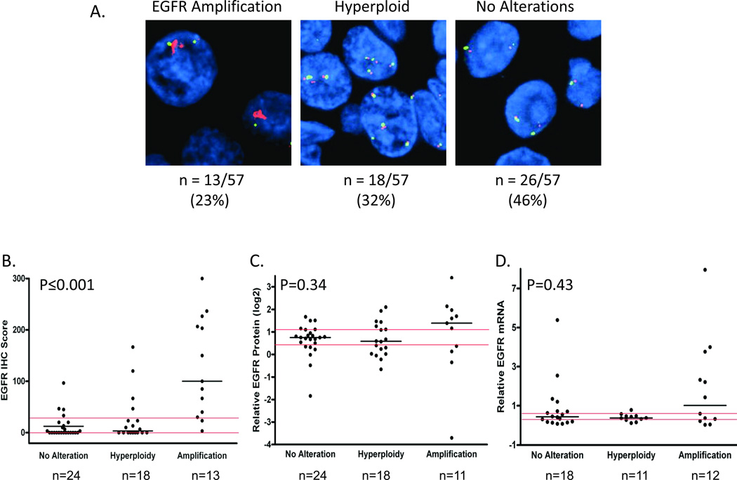
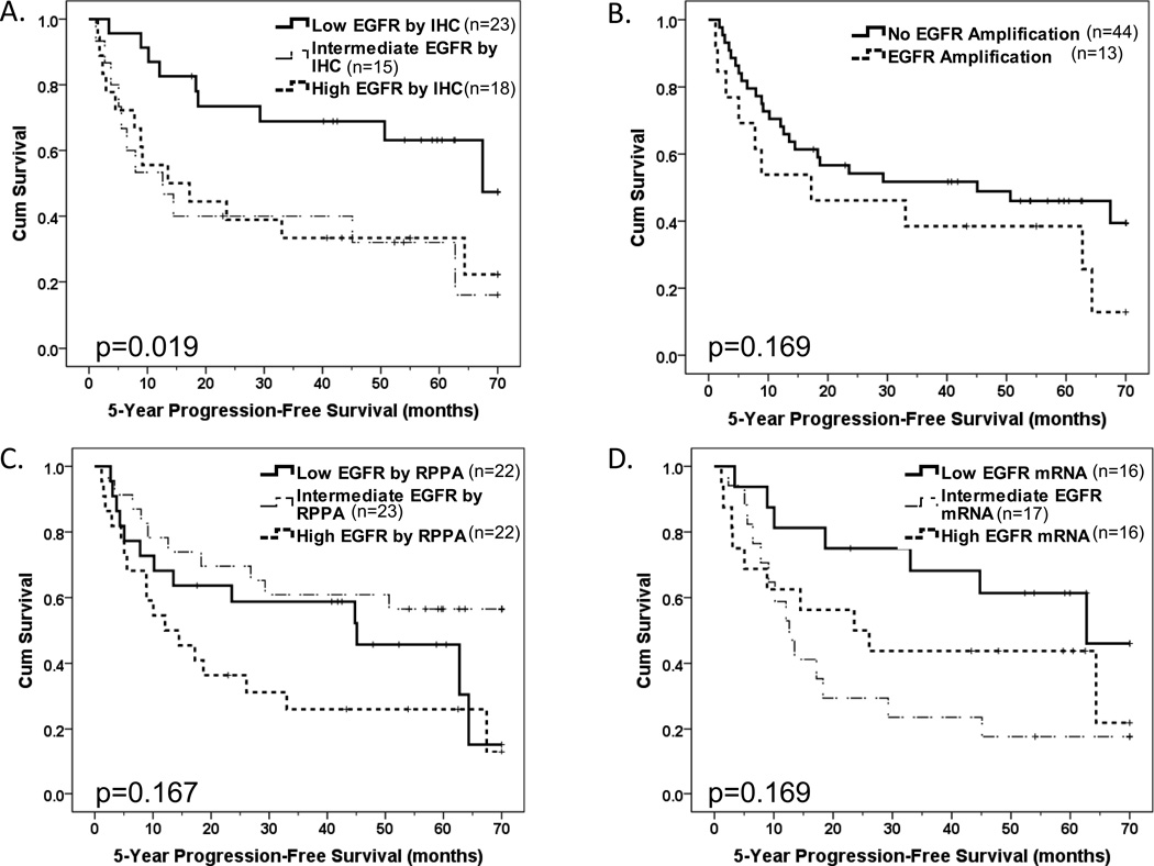
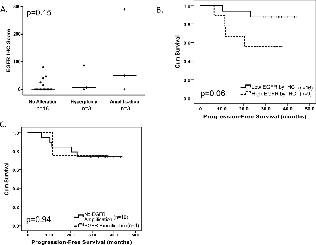
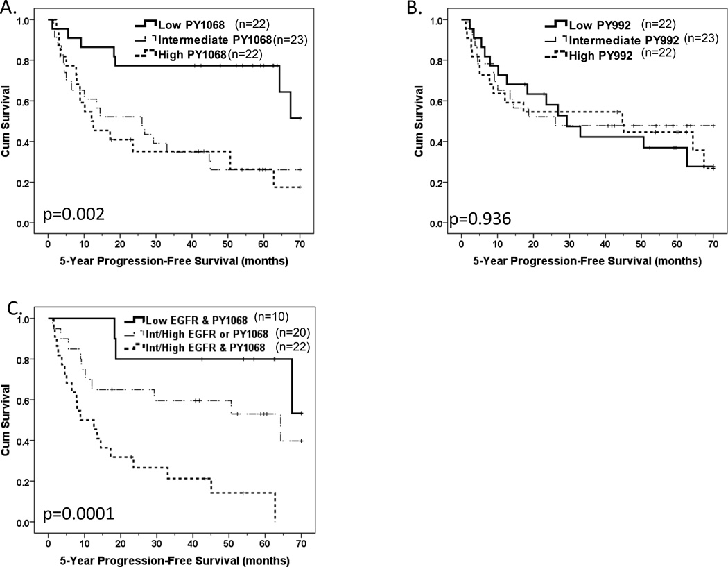
Patients and methods: HNSCC tumors from patients prospectively enrolled in either an Early Detection Research Network (EDRN) study and treated with surgery without an EGFR-targeted agent (N = 154) or enrolled in a chemoradiation trial involving the EGFR-targeted antibody cetuximab (N = 39) were evaluated for EGFR gene amplification by FISH and EGFR protein by immunohistochemical staining. Fresh-frozen tumors (EDRN) were also evaluated for EGFR protein and site-specific phosphorylation at Y992 and Y1068 using reverse-phase protein array (n = 67). Tumor (n = 50) EGFR and EGFRvIII mRNA levels were quantified using real-time PCR.
Results: EGFR expression by immunohistochemistry (IHC) was significantly higher in the EDRN tumors with EGFR gene amplification (P < 0.001), and a similar trend was noted in the cetuximab-treated cohort. In the EDRN and cetuximab-treated cohorts elevated EGFR by IHC was associated with reduced survival (P = 0.019 and P = 0.06, respectively). Elevated expression of total EGFR and EGFR PY1068 were independently significantly associated with reduced progression-free survival in the EDRN cohort [HR = 2.75; 95% confidence interval (CI) = 1.26-6.00 and HR = 3.29; 95% CI = 1.34-8.14, respectively].
Conclusions: In two independent HNSCC cohorts treated with or without cetuximab, tumor EGFR levels were indicative of survival. Tumor EGFR PY1068 levels provided prognostic information independent of total EGFR.
©2012 AACR.
Conflict of interest statement
Conflicts of Interest: The authors declare no conflicts of interest
Figures
References
-
- Altekruse SFKC, Krapcho M, Neyman N, Aminou R, Waldron W, Ruhl J, Howlader N, Tatalovich Z, Cho H, Mariotto A, Eisner MP, Lewis DR, Cronin K, Chen HS, Feuer EJ, Stinchcomb DG, Edwards BK, editors. SEER Cancer Statistics Review, 1975–2007. based on November 2009 SEER data submission. Bethesda, MD: National Cancer Institute; 2010.
-
- Conley BA. Treatment of advanced head and neck cancer: what lessons have we learned? J Clin Oncol. 2006;24:1023–1025. - PubMed
-
- Harris SL, Thorne LB, Seaman WT, Neil Hayes D, Couch ME, Kimple RJ. Association of p16(INK4a) overexpression with improved outcomes in young patients with squamous cell cancers of the oral tongue. Head Neck. 2010;33:1622–1627. - PubMed
-
- Muller S, Su L, Tighiouart M, Saba N, Zhang H, Shin DM, et al. Distinctive E-cadherin and epidermal growth factor receptor expression in metastatic and nonmetastatic head and neck squamous cell carcinoma: predictive and prognostic correlation. Cancer. 2008;113:97–107. - PubMed
-
- Chen B, van den Brekel MW, Buschers W, Balm AJ, van Velthuysen ML. Validation of tissue array technology in head and neck squamous cell carcinoma. Head Neck. 2003;25:922–930. - PubMed
Publication types
MeSH terms
Substances
Grants and funding
- K07 CA137140/CA/NCI NIH HHS/United States
- P30 CA016672/CA/NCI NIH HHS/United States
- CA16672/CA/NCI NIH HHS/United States
- P50CA097190/CA/NCI NIH HHS/United States
- P50 CA098258/CA/NCI NIH HHS/United States
- R01CA098372/CA/NCI NIH HHS/United States
- P50 CA097190/CA/NCI NIH HHS/United States
- F31 DE020223/DE/NIDCR NIH HHS/United States
- U01 CA084968/CA/NCI NIH HHS/United States
- U01CA84968/CA/NCI NIH HHS/United States
- R01 CA077308/CA/NCI NIH HHS/United States
- R01 CA098372/CA/NCI NIH HHS/United States
- K07CA137140/CA/NCI NIH HHS/United States
- 1F31DE020223-01A1/DE/NIDCR NIH HHS/United States
LinkOut - more resources
Full Text Sources
Other Literature Sources
Medical
Research Materials
Miscellaneous
