. 2012 Apr 13;287(16):13303-12.
doi: 10.1074/jbc.M111.294041. Epub 2012 Feb 20.

Ligation of the fibrin-binding domain by β-strand addition is sufficient for expansion of soluble fibronectin

Affiliations

Ligation of the fibrin-binding domain by β-strand addition is sufficient for expansion of soluble fibronectin

Lisa M Maurer et al. J Biol Chem. .

Abstract

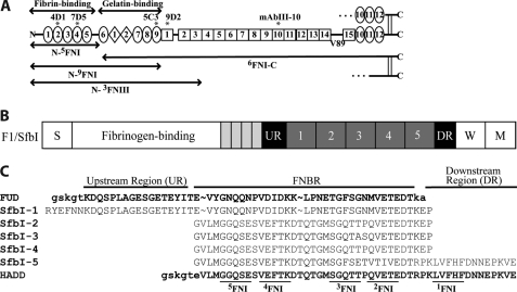
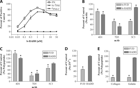
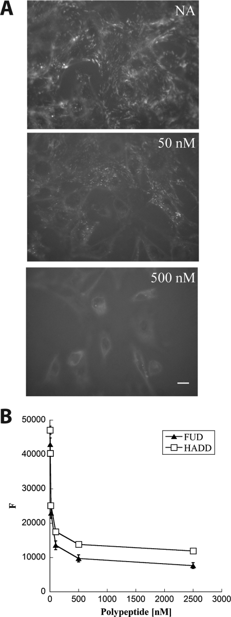
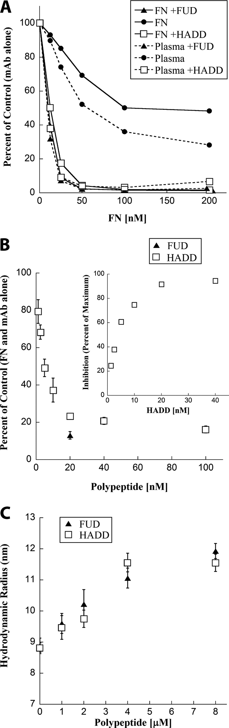
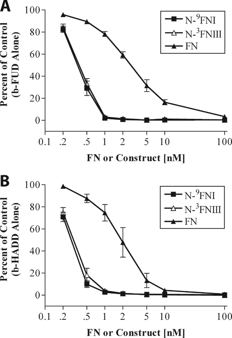
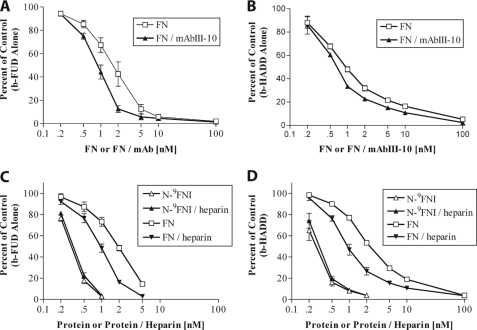
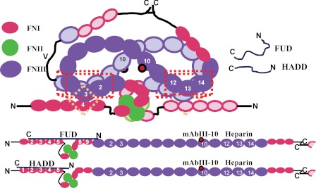
How fibronectin (FN) converts from a compact plasma protein to a fibrillar component of extracellular matrix is not understood. "Functional upstream domain" (FUD), a polypeptide based on F1 adhesin of Streptococcus pyogenes, binds by anti-parallel β-strand addition to discontinuous sets of N-terminal FN type I modules, (2-5)FNI of the fibrin-binding domain and (8-9)FNI of the gelatin-binding domain. Such binding blocks assembly of FN. To learn whether ligation of (2-5)FNI, (8-9)FNI, or the two sets in combination is important for inhibition, we tested "high affinity downstream domain" (HADD), which binds by β-strand addition to the continuous set of FNI modules, (1-5)FNI, comprising the fibrin-binding domain. HADD and FUD were similarly active in blocking fibronectin assembly. Binding of HADD or FUD to soluble plasma FN exposed the epitope to monoclonal antibody mAbIII-10 in the tenth FN type III module ((10)FNIII) and caused expansion of FN as assessed by dynamic light scattering. Soluble N-terminal constructs truncated after (9)FNI or (3)FNIII competed better than soluble FN for binding of FUD or HADD to adsorbed FN, indicating that interactions involving type III modules more C-terminal than (3)FNIII limit β-strand addition to (1-5)FNI within intact soluble FN. Preincubation of FN with mAbIII-10 or heparin modestly increased binding to HADD or FUD. Thus, ligation of FNIII modules involved in binding of integrins and glycosaminoglycans, (10)FNIII and (12-14)FNIII, increases accessibility of (1-5)FNI. Allosteric loss of constraining interactions among (1-5)FNI, (10)FNIII, and (12-14)FNIII likely enables assembly of FN into extracellular fibrils.


Figures

FIGURE 1.
FIGURE 1.
Diagram of FN and FN constructs, schematic of the F1 adhesin, and sequences of HADD versus FUD. A, each subunit of FN consists of 12 FNI modules (ovals), 2 FNII modules (diamonds), and 15 FNIII modules (squares) for the V89 splice variant shown. In plasma FN, one subunit contains a variable region, and the other subunit lacks it. Modules are numbered to facilitate naming recombinant proteins according to modular content. The boundaries of fibrin- and gelatin-binding domains are indicated, as are locations of epitopes for mAbs (asterisks). B, schematic of the F1 adhesin showing the signal sequence (S), four proline-rich repeats (small rectangles), upstream region (UR), numbered FNBRs, downstream region (DR), the wall-spanning region (W), and membrane-spanning region (M). C, five FNBRs and the upstream and downstream nonrepetitive regions of F1 adhesin (SfbI) and relationship of this sequence to FUD and HADD. The tails introduced into FUD or HADD by the cloning strategy are in lowercase. In HADD, 33 residues of the second or fourth FNBR are joined to 16 residues of the downstream region to add the binding sequence for 1FNI. The underlined sequences in HADD are predicted to interact with the indicated FNI modules (22, 24, 35).
FIGURE 2.
FIGURE 2.
HADD binds to FN via N-5FNI. A, enzyme-linked assay of increasing concentrations of biotinylated-HADD (b-HADD) binding to wells coated with 40 nm FN (□), N-5FNI (▴), or 6FNIII-C (▾). The amount bound was normalized to a positive control, and wells were coated with b-FUD at 1 μg/ml. B, binding relative to no mAb of 0.3 nm b-HADD or b-FUD to coated FN in the presence of 30 μg/ml 4D1 to 2FNI, 7D5 to 4FNI, or 5C3 to 9FNI. C, binding relative to no peptide of 4D1 (1:50,000 ascites), 7D5 (1:50,000 ascites), or 5C3 (1:30,000 ascites) in the presence of 175 nm HADD or FUD. D, binding relative to no Zn2+ of 0.3 nm b-HADD or b-FUD incubated with coated FN in the absence or presence of 1 mm Zn2+. E, binding of FN to collagen or gelatin in the presence or absence of 100 nm HADD or FUD as detected by 9D2. Values are mean ± S.D. of three (A–D) or two experiments. Significance of the differences from the indicated 100% controls were calculated by a t test. B–E, differences of p < 0.05 from 100% controls are indicated by asterisks.
FIGURE 3.
FIGURE 3.
HADD inhibits FN assembly by fibroblasts. A, mouse FN−/− cells adherent to laminin-coated coverslips were given 20 nm FITC-FN in the absence (NA) or presence of 50 or 500 nm HADD. Following incubation for 1 h, cells were washed, fixed, and imaged via fluorescence microscopy. Photomicrographs were taken at the exposure time determined for NA control and manipulated similarly. Bar, 10 μm. Shown are typical images from multiple fields in two different experiments. Similar results were seen in additional experiments not shown on FN−/− cells adherent to FN or to FN lacking the N-9FNI region. B, dose-dependent inhibition of A488-FN incorporation into fibroblast matrices by HADD or FUD. A488-FN (20 nm) in 2% calf serum was incubated for 18 h with monolayers of human foreskin fibroblasts in the presence or absence of the indicated concentrations of FUD or HADD. Following washes in PBS, fluorescence intensity (F) was measured in a microplate reader. In the experiment shown, the fluorescence of cells not treated with A488-FN was 3900 and subtracted from each of the values. The results are typical of four experiments done with FUD and two experiments done with HADD.
FIGURE 4.
FIGURE 4.
HADD is similar to FUD in exposing the mAbIII-10 epitope of purified FN or FN in plasma and expanding purified FN. A, effect of HADD or FUD on the exposure of the mAbIII-10 epitope in purified FN and FN in plasma as determined by competitive ELISA. Purified FNs (solid lines) or FNs in diluted plasma (dotted lines), 233 nm, were incubated without (●) or with 580 nm FUD (▴) or HADD (□) for 30 min. Prior to the assay, the concentration of FN in neat plasma was found to be 0.65 mg/ml (2600 nm) by competition ELISA with a mAb that is not conformation-sensitive. The six samples were then diluted to the indicated FN concentrations; mAbIII-10 was added, and competition by soluble FN for mAbIII-10 binding to coated FN was determined. Data are expressed as percent of mAbIII-10 binding alone (no FN or polypeptide added) and are representative of two experiments. B, ELISA of competition of binding of mAbIII-10 to coated FN by 20 nm soluble FN alone or preincubated with the indicated concentrations of HADD (□) or with 20 nm FUD (▴). Values are expressed relative to 20 nm soluble FN with mAbIII-10 but no polypeptide and represent mean ± S.D. of three experiments. The inset replots the HADD titration in comparison with the maximum inhibition found with 40 nm HADD. C, HADD (□) or FUD (▴) were titrated into FN solution separately, and the hydrodynamic radius of 4 μm FN or 4 μm FN plus the indicated concentration of polypeptide was calculated. Measurements were performed at 25 °C in 20 mm Tris, 100 mm sodium chloride, pH 7.4. Error bars indicate the standard deviation of six measurements on each sample. The experiment was repeated twice with the same result.
FIGURE 5.
FIGURE 5.
N-9FNI and N-3FNIII compete similarly for FUD or HADD binding to adsorbed FN. Binding of 0.3 nm b-FUD (A) or 0.3 nm b-HADD (B) to coated FN in the presence of increasing concentrations of soluble N-9FNI (■), N-3FNII (△), or FN (▴). Assays were in Tris buffer, pH 7.4, containing 50 mm NaCl. Values are expressed relative to biotinylated polypeptide alone and represent mean ± S.D. of three experiments.
FIGURE 6.
FIGURE 6.
Complex formation with mAbIII-10 or heparin increases competition by soluble FN for binding of FUD or HADD to adsorbed N-9FNI or FN. A and B, competition for binding of 0.3 nm b-FUD (A) or b-HADD (B) to coated N-9FNI by increasing concentrations of soluble FN (□) or FN plus mAbIII-10 (present at a ratio of 1 IgG per FN subunit) (▴). Assays were done in Tris buffer, pH 7.4, containing 300 mm NaCl. C and D, competition for binding of 0.3 nm b-FUD (C) or b-HADD (D) to coated FN in the presence of N-9FNI (△), N-9FNI plus 0.25 mg/ml heparin (▴), FN (□), or FN plus 0.25 mg/ml heparin (▾). Assays were done in Tris buffer, pH 7.4, containing 150 mm NaCl. Values are expressed relative to biotinylated polypeptide alone and represent mean ± S.D. of 3 (A), 2 (B), 3 (C), or 2–3 (D) experiments.
FIGURE 7.
FIGURE 7.
Diagram of how FUD and HADD may cause expansion of plasma FN. Plasma FN in a conceptual compact conformation (top) or extended conformation (bottom). One subunit is drawn with completely filled symbols, the other with outlined symbols. The complete dimer is shown in the compact conformation, and one subunit and part of the second is shown for the extended conformation. Only selected modules are numbered. The subunits are held together by disulfides at the C termini and interactions of 12–14FNIII with 2–3FNIII (red three-dimensional box). Each subunit is further constrained by the 4FNI-3FNIII interaction (pink diamond). FUD or HADD, which by themselves are random coils, form β-zippers with the indicated FNI modules, resulting in unfolding and expansion of the quaternary structure. The epitope for mAbIII-10 used to monitor conformational change is in 10FNIII (red dot).

Similar articles

Cited by

References

    1. Pankov R., Yamada K. M. (2002) Fibronectin at a glance. J. Cell Sci. 115, 3861–3863 - PubMed
    1. Tucker R. P., Chiquet-Ehrismann R. (2009) Evidence for the evolution of tenascin and fibronectin early in the chordate lineage. Int. J. Biochem. Cell Biol. 41, 424–434 - PubMed
    1. Maurer L. M., Tomasini-Johansson B. R., Mosher D. F. (2010) Emerging roles of fibronectin in thrombosis. Thromb. Res. 125, 287–291 - PMC - PubMed
    1. Odermatt E., Engel J. (1989) Physical Properties of Fibronectin in Fibronectin (Mosher D. F., ed) pp. 29–45, Academic Press, Inc., San Diego, CA
    1. Rocco M., Infusini E., Daga M. G., Gogioso L., Cuniberti C. (1987) Models of fibronectin. EMBO J. 6, 2343–2349 - PMC - PubMed

Publication types

LinkOut - more resources