. 2012 Apr 13;287(16):13128-36.
doi: 10.1074/jbc.M111.315788. Epub 2012 Feb 18.

Phosphatidylinositol 5-phosphatase oculocerebrorenal syndrome of Lowe protein (OCRL) controls actin dynamics during early steps of Listeria monocytogenes infection

Affiliations

Phosphatidylinositol 5-phosphatase oculocerebrorenal syndrome of Lowe protein (OCRL) controls actin dynamics during early steps of Listeria monocytogenes infection

Andreas Kühbacher et al. J Biol Chem. .

Abstract

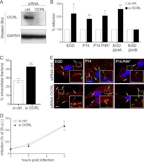
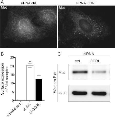
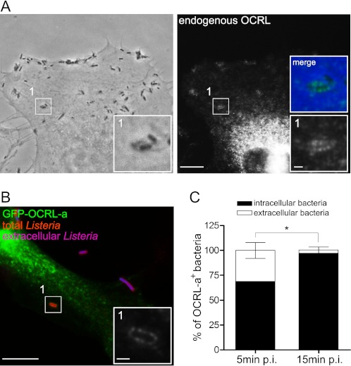
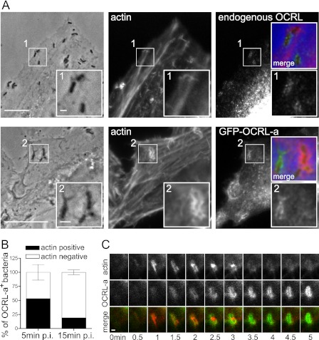
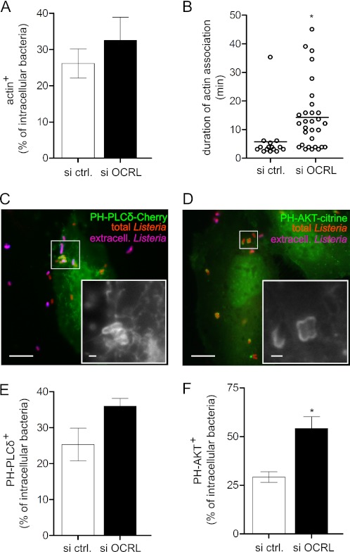
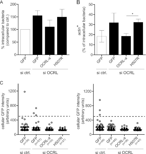
Listeria monocytogenes is a bacterial pathogen that induces its own entry into a broad range of mammalian cells through interaction of the bacterial surface protein InlB with the cellular receptor Met, promoting an actin polymerization/depolymerization process that leads to pathogen engulfment. Phosphatidylinositol bisphosphate (PI[4,5]P(2)) and trisphosphate (PI[3,4,5]P(3)) are two major phosphoinositide species that function as molecular scaffolds, recruiting cellular effectors that regulate actin dynamics during L. monocytogenes infection. Because the phosphatidylinositol 5'-phosphatase OCRL dephosphorylates PI(4,5)P(2) and to a lesser extent PI(3,4,5)P(3), we investigated whether this phosphatase modulates cell invasion by L. monocytogenes. Inactivation of OCRL by small interfering RNA (siRNA) leads to an increase in the internalization levels of L. monocytogenes in HeLa cells. Interestingly, OCRL depletion does not increase but rather decreases the surface expression of the receptor Met, suggesting that OCRL controls bacterial internalization by modulating signaling cascades downstream of Met. Immuno-fluorescence microscopy reveals that endogenous and overexpressed OCRL are present at L. monocytogenes invasion foci; live-cell imaging additionally shows that actin depolymerization coincides with EGFP-OCRL-a accumulation around invading bacteria. Together, these observations suggest that OCRL promotes actin depolymerization during L. monocytogenes infection; in agreement with this hypothesis, OCRL depletion leads to an increase in actin, PI(4,5)P(2), and PI(3,4,5)P(3) levels at bacterial internalization foci. Furthermore, in cells knocked down for OCRL, transfection of enzymatically active EGFP-OCRL-a (but not of a phosphatase-dead enzyme) decreases the levels of intracellular L. monocytogenes and of actin associated with invading bacteria. These results demonstrate that through its phosphatase activity, OCRL restricts L. monocytogenes invasion by modulating actin dynamics at bacterial internalization sites.


Figures

FIGURE 1.
FIGURE 1.
OCRL depletion promotes L. monocytogenes invasion of HeLa cells. A, OCRL expression was knocked down in HeLa cells for 72 h using specific siRNA oligonucleotides, and protein levels were verified by Western blot employing GAPDH as a loading control. siRNA ctrl., siRNA control. B, gentamicin invasion assays were performed in HeLa cells transfected with control or anti-OCRL siRNA using five different L. monocytogenes strains: EGD, P14, P14.PrfA*, EGD ΔInlA, and EGD ΔInlB. Values represent the averages ± S.E. of tetraplicate samples from 3–4 independent experiments (Student's t test: *, p < 0.05 and **, p < 0.01). C, HeLa cells transfected with control or anti-OCRL siRNA were infected for 1 h with L. monocytogenes P14.PrfA* strain, and cells were processed for immuno-fluorescence microscopy with specific labeling of total versus extracellular bacteria. Values represent the averages ± S.E. of 3 independent experiments (n = 200 bacteria analyzed per experiment; Student's t test: **, p < 0.01). D, gentamicin invasion assays for L. monocytogenes EGD were done in OCRL-depleted and control cells at 2, 3, and 5 h after infection to monitor intracellular growth. Values represent the averages ± S.E. of tetraplicate samples from one representative experiment out of two independent experiments. p.i., post-infection. E, cells knocked down for OCRL or transfected with control siRNA were infected with the indicated L. monocytogenes strains. Cells were processed for immuno-fluorescence. DNA was labeled with Hoechst (blue), L. monocytogenes were labeled with antibodies against the bacterial cell wall (green), and actin comet tails polymerized by intracellular bacteria (arrows) were labeled with fluorescent phalloidin (red). Bar: 10 μm (inset bar: 1 μm).
FIGURE 2.
FIGURE 2.
OCRL depletion decreases expression of Met. A, after 72 h of transfection with control or anti-OCRL siRNA, HeLa cells were fixed and processed for immuno-fluorescence microscopy in saponin permeabilized cells using the anti-Met C-12 antibody. Bar: 10 μm. siRNA ctrl., siRNA control. B, surface expression of Met in control and OCRL-depleted cells was quantified by flow cytometry without cellular permeabilization using the DO-24 antibody against the extracellular moiety of Met. The results represents the -fold change of geometric mean as compared with unstained cells ± S.E. of 3 independent experiments (Student's t test: **, p < 0.01). C, expression of Met in control and OCRL-depleted cells was verified by Western blot using the anti-Met C-12 antibody.
FIGURE 3.
FIGURE 3.
OCRL is recruited to L. monocytogenes internalization sites. A, HeLa cells were infected for 10 min with L. monocytogenes P14.PrfA* and processed for immuno-fluorescence microscopy by labeling endogenous OCRL. Bar: 10 μm (inset bar: 1 μm). B, HeLa cells were transfected for 24 h with EGFP-OCRL-a (green) and were subsequently infected for 10 min with L. monocytogenes P14.PrfA*; cells were then processed for fluorescence microscopy with differential labeling of extracellular (blue) and total bacteria (red). The inset highlights a specific area of the EGFP-OCRL-a channel. Bar: 10 μm (inset bar: 1 μm). C, quantification of the proportion of intracellular versus extracellular bacteria that were labeled by the EGFP-OCRL-a marker in cells processed for B. Values represent the averages ± S.E. of 3 independent experiments (Student's t test: *, p < 0.05). p.i., post-infection.
FIGURE 4.
FIGURE 4.
Actin polymerization precedes OCRL recruitment to L. monocytogenes vacuoles. A, microscopy analysis of the distribution of endogenous OCRL, EGFP-OCRL-a, and actin in cells infected by L. monocytogenes P14.PrfA*. Bar: 10 μm (inset bar: 1 μm). B, quantification of the recruitment of OCRL and actin by L. monocytogenes P14.PrfA*. Values represent the averages ± S.E. of 3 independent experiments. p.i., post-infection. C, live-cell imaging illustrating the sequential recruitment of actin visualized by LifeAct-cherry and OCRL by L. monocytogenes P14.PrfA*. Bar: 1 μm.
FIGURE 5.
FIGURE 5.
OCRL depletion increases association of actin, PI(4,5)P2 and PI(3,4,5)P3 to L. monocytogenes vacuoles. A, association between actin and intracellular L. monocytogenes P14.PrfA* on fixed cells 15 min after infection. Values represent the averages ± S.E. of 4 independent experiments. si ctrl., siRNA control. B, time of association of single bacteria to actin visualized by LifeAct-cherry in live-cell imaging experiments; horizontal bars indicate the arithmetic mean (Student's t test: *, p < 0.05). C, association between PI(4,5)P2 visualized by PH-PLC-δ-3-cherry and L. monocytogenes P14.PrfA* on fixed cells 25 min after infection. Bar: 10 μm (inset bar: 1 μm). D, association between PI (3,4,5)P3 visualized by PH-AKT-citrine and L. monocytogenes P14.PrfA* on fixed cells 25 min after infection. Bar: 10 μm (inset bar: 1 μm). E, quantification of PH-PLCδ-3-cherry association to intracellular L. monocytogenes in fixed cells 25 min after infection. F, quantification of PH-AKT-citrine association to intracellular L. monocytogenes in fixed cells 25 min after infection Values represent the averages ± S.E. of 3 independent experiments (Student's t test: *, p < 0.05).
FIGURE 6.
FIGURE 6.
OCRL phosphatase function is responsible for restricting L. monocytogenes invasion and for promoting actin depolymerization on bacterial vacuoles. A, one set of HeLa cells was transfected with control siRNA and a plasmid coding for GFP, whereas a second set of HeLa cells was transfected with anti-OCRL siRNA and plasmids coding for GFP, siRNA-resistant pEGFP-OCRL-a, or pEGFP-OCRL-a phosphatase-dead (H507R). After 1 h of infection with L. monocytogenes P14.PrfA*, cells were processed for immuno-fluorescence, and invasion was quantified by scoring extracellular versus total cell-associated bacteria. Values represent the averages ± S.E. of 3 independent experiments. si ctrl., siRNA control. B, transfections were performed as described in A, and after 15 min of infection with L. monocytogenes P14.PrfA*, cells were processed for immuno-fluorescence microscopy to quantify the percentage of intracellular bacteria associated to actin (labeled by fluorescent phalloidin). Values represent the averages ± S.E. of 3 independent experiments. (Student's t test: *, p < 0.05.) C, the GFP fluorescence intensity of cells chosen for quantifications in A and B was measured using ImageJ. Circles represent cells, and horizontal lines represent the average of all cells of the given condition. For quantification, low to medium GFP-expressing cells with a GFP intensity lower than 500 units (dashed line) were considered.

References

    1. Cossart P, (2011) Illuminating the landscape of host-pathogen interactions with the bacterium Listeria monocytogenes. Proc. Natl. Acad. Sci. U.S.A. 108, 19484–19491 - PMC - PubMed
    1. Stavru F., Archambaud C., Cossart P. (2011) Cell biology and immunology of Listeria monocytogenes infections: novel insights. Immunol. Rev. 240, 160–184 - PubMed
    1. Mengaud J., Ohayon H., Gounon P., Mege R.-M., Cossart P. (1996) E-cadherin is the receptor for internalin, a surface protein required for entry of L. monocytogenes into epithelial cells. Cell 84, 923–932 - PubMed
    1. Shen Y., Naujokas M., Park M., Ireton K. (2000) InIB-dependent internalization of Listeria is mediated by the Met receptor tyrosine kinase. Cell 103, 501–510 - PubMed
    1. Pizarro-Cerdá J., Cossart P. (2009) Listeria monocytogenes membrane trafficking and lifestyle: the exception or the rule? Annu. Rev. Cell Dev. Biol. 25, 649–670 - PubMed

Publication types

MeSH terms