

Splice-specific glycine receptor binding, folding, and phosphorylation of the scaffolding protein gephyrin
- PMID: 22351777
- PMCID: PMC3339950
- DOI: 10.1074/jbc.M112.341826
Splice-specific glycine receptor binding, folding, and phosphorylation of the scaffolding protein gephyrin
Abstract
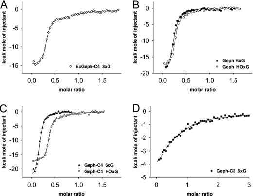
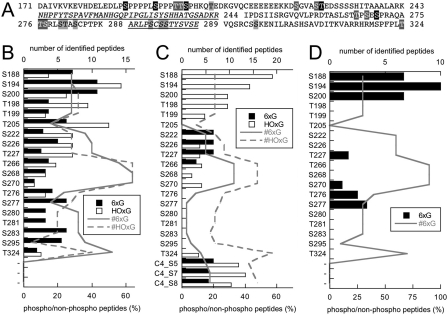
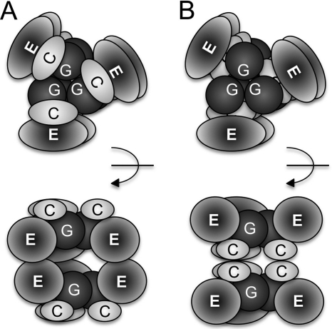
The multimeric scaffolding protein gephyrin forms post-synaptic clusters at inhibitory sites, thereby anchoring inhibitory glycine (GlyR) and subsets of γ-aminobutyric acid type A (GABAA) receptors. Gephyrin is composed of three domains, the conserved N-terminal G- and C-terminal E-domain, connected by the central (C-) domain. In this study we investigated the oligomerization, folding and stability, GlyR β-loop binding, and phosphorylation of three gephyrin splice variants (Geph, Geph-C3, Geph-C4) after expression and purification from insect cells (Sf9). In contrast to Escherichia coli-derived trimeric gephyrin, we found that Sf9 gephyrins form hexamers as basic oligomeric form. In the case of Geph and Geph-C4, also high-oligomeric forms (∼900 kDa) were isolated. Partial proteolysis revealed a compact folding of the Gephyrin G and C domain in one complex, whereas a much lower stability for the E domain was found. After GlyR β-loop binding, the stability of the E domain increased in Geph and Geph-C4 significantly. In contrast, the E domain in Geph-C3 is less stable and binds the GlyR β-loop with one order of magnitude lower affinity. Finally, we identified 18 novel phosphorylation sites in gephyrin, of which all except one are located within the C domain. We propose two models for the domain arrangement in hexameric gephyrin based on the oligomerization of either the E or C domains, with the latter being crucial for the regulation of gephyrin clustering.
Figures
Similar articles
-
Regulation of gephyrin assembly and glycine receptor synaptic stability.J Biol Chem. 2006 Oct 6;281(40):30046-56. doi: 10.1074/jbc.M602155200. Epub 2006 Aug 1. J Biol Chem. 2006. PMID: 16882665
-
A gephyrin-related mechanism restraining glycine receptor anchoring at GABAergic synapses.J Neurosci. 2004 Feb 11;24(6):1398-405. doi: 10.1523/JNEUROSCI.4260-03.2004. J Neurosci. 2004. PMID: 14960612 Free PMC article.
-
Crystal structures of human gephyrin and plant Cnx1 G domains: comparative analysis and functional implications.J Mol Biol. 2001 Sep 14;312(2):405-18. doi: 10.1006/jmbi.2001.4952. J Mol Biol. 2001. PMID: 11554796
-
Gephyrin and the regulation of synaptic strength and dynamics at glycinergic inhibitory synapses.Brain Res Bull. 2017 Mar;129:50-65. doi: 10.1016/j.brainresbull.2016.09.003. Epub 2016 Sep 6. Brain Res Bull. 2017. PMID: 27612963 Review.
-
Gephyrin: where do we stand, where do we go?Trends Neurosci. 2008 May;31(5):257-64. doi: 10.1016/j.tins.2008.02.006. Epub 2008 Apr 9. Trends Neurosci. 2008. PMID: 18403029 Review.
Cited by
-
Gephyrin: a key regulatory protein of inhibitory synapses and beyond.Histochem Cell Biol. 2018 Nov;150(5):489-508. doi: 10.1007/s00418-018-1725-2. Epub 2018 Sep 27. Histochem Cell Biol. 2018. PMID: 30264265 Review.
-
Nanoscale Subsynaptic Domains Underlie the Organization of the Inhibitory Synapse.Cell Rep. 2019 Mar 19;26(12):3284-3297.e3. doi: 10.1016/j.celrep.2019.02.070. Cell Rep. 2019. PMID: 30893601 Free PMC article.
-
Thermodynamic modulation of gephyrin condensation by inhibitory synapse components.Proc Natl Acad Sci U S A. 2024 Mar 19;121(12):e2313236121. doi: 10.1073/pnas.2313236121. Epub 2024 Mar 11. Proc Natl Acad Sci U S A. 2024. PMID: 38466837 Free PMC article.
-
The biosynthesis of the molybdenum cofactors.J Biol Inorg Chem. 2015 Mar;20(2):337-47. doi: 10.1007/s00775-014-1173-y. Epub 2014 Jul 1. J Biol Inorg Chem. 2015. PMID: 24980677
-
An aggregation-removal model for the formation and size determination of post-synaptic scaffold domains.PLoS Comput Biol. 2017 Apr 24;13(4):e1005516. doi: 10.1371/journal.pcbi.1005516. eCollection 2017 Apr. PLoS Comput Biol. 2017. PMID: 28437460 Free PMC article.
References
-
- Fritschy J. M., Harvey R. J., Schwarz G. (2008) Gephyrin. Where do we stand, where do we go? Trends Neurosci. 31, 257–264 - PubMed
-
- Kneussel M., Hermann A., Kirsch J., Betz H. (1999) Hydrophobic interactions mediate binding of the glycine receptor β-subunit to gephyrin. J. Neurochem. 72, 1323–1326 - PubMed
-
- Schrader N., Kim E. Y., Winking J., Paulukat J., Schindelin H., Schwarz G. (2004) Biochemical characterization of the high affinity binding between the glycine receptor and gephyrin. J. Biol. Chem. 279, 18733–18741 - PubMed
Publication types
MeSH terms
Substances
LinkOut - more resources
Full Text Sources
Miscellaneous
