. 2012 Mar 30;287(14):11141-50.
doi: 10.1074/jbc.M111.330209. Epub 2012 Feb 17.

CutA divalent cation tolerance homolog (Escherichia coli) (CUTA) regulates β-cleavage of β-amyloid precursor protein (APP) through interacting with β-site APP cleaving protein 1 (BACE1)

Affiliations

CutA divalent cation tolerance homolog (Escherichia coli) (CUTA) regulates β-cleavage of β-amyloid precursor protein (APP) through interacting with β-site APP cleaving protein 1 (BACE1)

Yingjun Zhao et al. J Biol Chem. .

Abstract

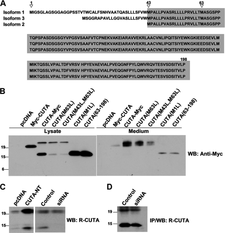
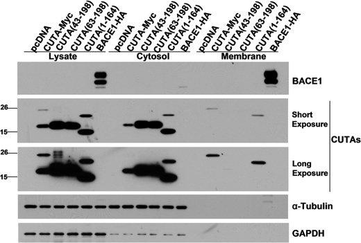
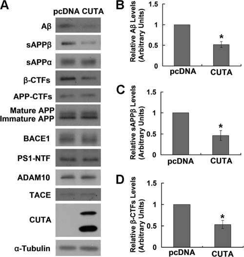
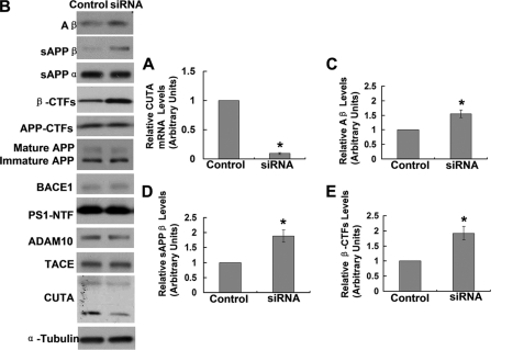
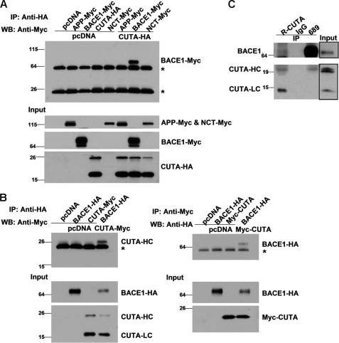
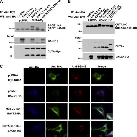
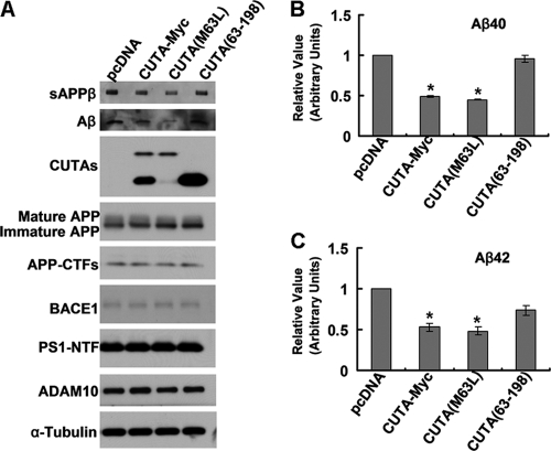
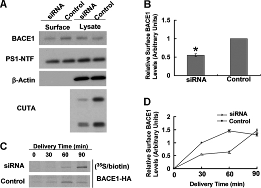
Accumulation of the neurotoxic β-amyloid (Aβ) peptide in the brain is central to the pathogenesis of Alzheimer disease. Aβ is derived from the β-amyloid precursor protein (APP) through sequential cleavages by β- and γ-secretases, and the production of Aβ is greatly affected by the subcellular localization of these factors. CUTA, the mammalian CutA divalent cation tolerance homolog (E. coli), has been proposed to mediate acetylcholinesterase activity and copper homeostasis, which are important in Alzheimer disease pathology. However, the exact function of CUTA remains largely unclear. Here we show that human CUTA has several variants that differ in their N-terminal length and are separated as heavy (H) and light (L) components. The H component has the longest N terminus and is membrane-associated, whereas the L component is N-terminally truncated at various sites and localized in the cytosol. Importantly, we demonstrate that the H component of CUTA interacts through its N terminus with the transmembrane domain of β-site APP cleaving enzyme 1 (BACE1), the putative β-secretase, mainly in the Golgi/trans-Golgi network. Overexpression and RNA interference knockdown of CUTA can reduce and increase BACE1-mediated APP processing/Aβ secretion, respectively. RNA interference of CUTA decelerates intracellular trafficking of BACE1 from the Golgi/trans-Golgi network to the cell surface and reduces the steady-state level of cell surface BACE1. Our results identify the H component of CUTA as a novel BACE1-interacting protein that mediates the intracellular trafficking of BACE1 and the processing of APP to Aβ.


Figures

FIGURE 1.
FIGURE 1.
Human CUTA has several variants that can be separated into heavy and light components. A, alignment of the three known human CUTA isoforms is shown. Identical amino acids shared by the three isoforms are highlighted in gray. Potential translation initiation methionine sites are indicated by triangles. B, HEK 293T cells were transfected with control pcDNA, N-terminal tagged CUTA (Myc-CUTA), C-terminal tagged CUTA (CUTA-Myc), C-terminal Myc-tagged CUTA with methionine 1 mutated to leucine (M1L), methionine 63 mutated to leucine (M63L), or methionines 43 and 63 mutated to leucines (M43L, M63L), and C-terminal Myc-tagged CUTA that had amino acids 1–62 of its N terminus truncated (63–198). After 44 h, culture media was changed to fresh media. After another 4 h, conditioned media was collected, and cells were lysed. Equal protein amounts of cell lysates and equal volume amounts of conditioned media were subjected to SDS-PAGE and Western blot (WB) using the Myc antibody 9E10. C, cells were transfected with pcDNA or non-tagged CUTA (CUTA-NT) (left panel) or transfected with control siRNA or CUTA siRNA 3 (right panel). Equal protein amounts of cell lysates were subjected to SDS-PAGE and Western blot using the R-CUTA antibody. D, lysates containing equalized amounts of protein from cells transfected with control siRNA or CUTA siRNA 3 were incubated with the R-CUTA antibody for immunoprecipitation (IP). Immunoprecipitated proteins were analyzed by Western blot using the same antibody.
FIGURE 2.
FIGURE 2.
The heavy component of CUTA containing the N terminus is associated with the membrane, whereas the light component of CUTA is located in the cytosol. Cells were transfected with pcDNA, full-length CUTA, CUTA with N-terminal amino acids 1–42 truncated (43–198), CUTA with N-terminal amino acids 1–62 truncated (63–198), CUTA with C-terminal amino acids 165–198 truncated (1–164), or BACE1-HA. After homogenization of cells, a small sample was used to analyze total lysates, and the rest was subjected to centrifuge to separate the cytosolic fraction and the membrane. The membrane pellet was rinsed with NaHCO3, pH 11.3, to dissociate peripheral membrane proteins and then resuspended in lysis buffer in a volume equal to that of the cytosolic fraction. Equal volume amounts of samples were subjected to SDS-PAGE and Western blot to detect CUTA (using the Myc antibody 9E10), GAPDH (to indicate membrane-bound protein), BACE1 (using an HA antibody to indicate the membrane fraction) and α-tubulin (to indicate the cytosolic fraction).
FIGURE 3.
FIGURE 3.
Overexpression of CUTA reduces β-processing of APP. A, HEK-Swe cells were transfected with control pcDNA and CUTA-Myc. The sAPPα and sAPPβ in conditioned media were Western-blotted with respective antibodies. Aβ secreted in conditioned media was precipitated by trichloroacetic acid and Western-blotted with 6E10. Cell lysates were Western-blotted with antibodies against total APP (mature and immature forms) and APP-CTFs (369), β-CTFs (6E10), BACE1 (3D5), PS1 NTF (Ab14), ADAM10, TACE, CUTA (Myc), and α-tubulin. The levels of Aβ (B), sAPPβ (C), and APP β-CTFs (D) were quantified by densitometry and normalized to those of controls for comparison (set as one arbitrary unit). *, p < 0.05, n = 3.
FIGURE 4.
FIGURE 4.
Down-regulation of CUTA increases β-processing of APP. HEK-Swe cells were transfected with control siRNA and CUTA siRNA 3. A, cells were subjected to RNA extraction and gene analysis by RT-PCR. The mRNA level of CUTA was normalized to that of GAPDH for comparison. *, p < 0.05, n = 3. B, the sAPPα and sAPPβ in conditioned media were Western-blotted with respective antibodies. Aβ secreted in conditioned media was precipitated by trichloroacetic acid and Western-blotted with 6E10. Cell lysates were Western-blotted with antibodies against total APP (mature and immature forms) and APP-CTFs (369), BACE1 (3D5), PS1 NTF (Ab14), ADAM10, TACE, CUTA (Myc), and α-tubulin. The levels of Aβ (C), sAPPβ (D), and APP β-CTFs (E) were quantified by densitometry and normalized to those of controls for comparison (set as one arbitrary unit). *, p < 0.05, n = 3.
FIGURE 5.
FIGURE 5.
The H component of CUTA interacts with BACE1. A, CUTA with an HA tag at the C terminus (CUTA-HA) was co-transfected with APP, BACE1, or NCT, all of which were Myc-tagged at the C terminus into HEK 293T cells. Cells co-transfected with pcDNA, and the indicated vectors were used as controls. Equal protein amounts of cell lysates were used for immunoprecipitation (IP) with an HA antibody and Western blot (WB) with the Myc antibody 9E10 to detect CUTA-interacting proteins. Ten percent of cell lysates were used as input to detect the expression of transfected proteins. B, pcDNA, BACE1-HA, and CUTA-Myc (left panel) or Myc-CUTA (right panel) were pairwise co-transfected into HEK 293T cells. Equal protein amounts of cell lysates were immunoprecipitated with an HA antibody and Western-blotted with a Myc antibody (left panels) or immunoprecipitated with the Myc antibody and Western-blotted with the HA antibody (right panels). Ten percent of cell lysates were used as input. *, nonspecific bands. C, the lysates of human brain tissues were immunoprecipitated with normal rabbit IgG, the R-CUTA antibody, or the BACE1 antibody 689. Immunoprecipitated proteins and input (5% of cell lysates) were subjected to SDS-PAGE and Western blot using another anti-BACE1 antibody (3D5) or R-CUTA.
FIGURE 6.
FIGURE 6.
The N terminus of CUTA and the transmembrane domain of BACE1 are crucial for their interaction in the Golgi/TGN. A, CUTA-Myc was co-transfected with BACE1, BACE1 lacking the C terminus (BACE1-ΔC), or BACE1 that has its transmembrane domain substituted with that of Nicastrin (BACE1(NCT/TM)), all of which were HA-tagged at the C terminus, into HEK 293T cells. Cells co-transfected with pcDNA and the indicated vectors were used as controls. Equal protein amounts of cell lysates were used for immunoprecipitation (IP) with a Myc antibody and Western blot (WB) with an HA antibody. Ten percent of cell lysates were used as input. B, BACE1-HA was co-transfected with CUTA, CUTA-(43–198), CUTA-(63–198), or CUTA-(1–164), all of which were Myc-tagged at the C terminus into HEK 293T cells. Equal protein amounts of cell lysates were used for IP with an HA antibody and Western blot with a Myc antibody. Ten percent of cell lysates were used as input. *, nonspecific bands. C, HeLa cells were transfected with pcDNA, Myc-CUTA, CUTA-(63–198), and/or BACE1-HA, as indicated. Cells were then fixed, permeabilized, and immunostained with primary antibodies against HA (indicating BACE1, in blue), Myc (indicating CUTA, in green), and TGN46 (indicating the Golgi/TGN organelle, in red), and respective secondary antibodies conjugated with Alexa Fluor 350, 488, or 594. Samples were examined by immunofluorescence microscopy.
FIGURE 7.
FIGURE 7.
Only BACE1-interacting CUTA forms regulate β-processing of APP and Aβ secretion. HEK-Swe cells were transfected with control pcDNA, CUTA-Myc, CUTA(M63L), or CUTA-(63–198). A, secreted Aβ and sAPPβ in conditioned media and mature/immature APP, APP-CTFs, BACE1, PS1-NTF, ADAM10, CUTA, and α-tubulin in cell lysates were detected by Western blot with respective antibodies. Secreted Aβ40 (B) and Aβ42 (C) in conditioned media were quantified by ELISA. The levels of Aβ40 and Aβ42 were normalized to those of controls for comparison (set as one arbitrary unit). *, p < 0.05, n = 4.
FIGURE 8.
FIGURE 8.
Down-regulation of CUTA slows cell surface delivery of BACE1 and reduces the steady-state level of BACE1 at the cell surface. A, after RNAi down-regulation of CUTA, live SH-SY5Y cells were subjected to biotinylation. Cell lysates were affinity-precipitated with streptavidin-agarose beads. The levels of biotinylated BACE1 and PS1-NTF as well as their total protein levels were analyzed by Western blot. B, cell surface levels of BACE1 in A were quantified by densitometry and normalized to that of a control (set as one arbitrary unit) for comparison. *, p < 0.05, n = 3. C, upon down-regulation of CUTA by RNAi, HEK 293T cells transfected with BACE1-HA were pulse-labeled with [35S]methionine for 15 min and incubated at 20 °C to accumulate isotope-labeled proteins in the TGN. Cells were then incubated at 37 °C for the indicated periods followed by biotinylation at 4 °C. After affinity precipitation, biotinylated proteins were immunoprecipitated with an anti-HA antibody and then subjected to SDS-PAGE and autoradiography. D, the band intensity in C was quantified by densitometry for comparison.

References

    1. Glenner G. G., Wong C. W. (1984) Alzheimer disease. Initial report of the purification and characterization of a novel cerebrovascular amyloid protein. Biochem. Biophys. Res. Commun. 120, 885–890 - PubMed
    1. Buée L., Bussière T., Buée-Scherrer V., Delacourte A., Hof P. R. (2000) Tau protein isoforms, phosphorylation, and role in neurodegenerative disorders. Brain Res. Brain Res. Rev. 33, 95–130 - PubMed
    1. Zhang Y. W., Thompson R., Zhang H., Xu H. (2011) APP processing in Alzheimer disease. Mol. Brain 4, 3. - PMC - PubMed
    1. Sinha S., Anderson J. P., Barbour R., Basi G. S., Caccavello R., Davis D., Doan M., Dovey H. F., Frigon N., Hong J., Jacobson-Croak K., Jewett N., Keim P., Knops J., Lieberburg I., Power M., Tan H., Tatsuno G., Tung J., Schenk D., Seubert P., Suomensaari S. M., Wang S., Walker D., Zhao J., McConlogue L., John V. (1999) Purification and cloning of amyloid precursor protein β-secretase from human brain. Nature 402, 537–540 - PubMed
    1. Vassar R., Bennett B. D., Babu-Khan S., Kahn S., Mendiaz E. A., Denis P., Teplow D. B., Ross S., Amarante P., Loeloff R., Luo Y., Fisher S., Fuller J., Edenson S., Lile J., Jarosinski M. A., Biere A. L., Curran E., Burgess T., Louis J. C., Collins F., Treanor J., Rogers G., Citron M. (1999) β-Secretase cleavage of Alzheimer amyloid precursor protein by the transmembrane aspartic protease BACE. Science 286, 735–741 - PubMed

Publication types

MeSH terms