. 2012 Mar 21;23(3):596-603.
doi: 10.1021/bc200647q. Epub 2012 Mar 1.

Dual 19F/1H MR gene reporter molecules for in vivo detection of β-galactosidase

Affiliations

Dual 19F/1H MR gene reporter molecules for in vivo detection of β-galactosidase

Jian-Xin Yu et al. Bioconjug Chem. .

Abstract

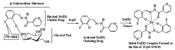
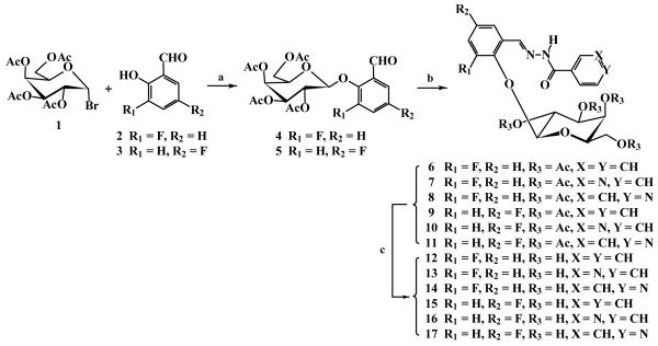
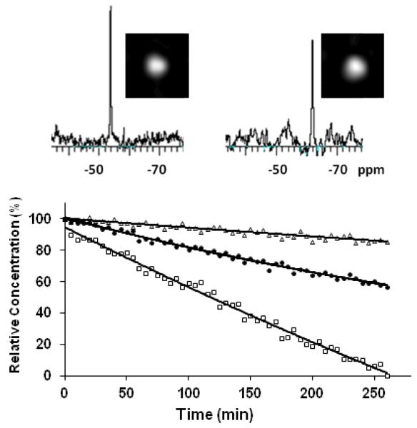
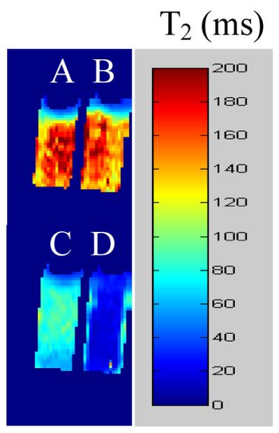
Increased emphasis on personalized medicine and novel therapies requires the development of noninvasive strategies for assessing biochemistry in vivo. The detection of enzyme activity and gene expression in vivo is potentially important for the characterization of diseases and gene therapy. Magnetic resonance imaging (MRI) is a particularly promising tool, since it is noninvasive and has no associated radioactivity, yet penetrates deep tissue. We now demonstrate a novel class of dual (1)H/(19)F nuclear magnetic resonance (NMR) lacZ gene reporter molecule to specifically reveal enzyme activity in human tumor xenografts growing in mice. We report the design, synthesis, and characterization of six novel molecules and evaluation of the most effective reporter in mice in vivo. Substrates show a single (19)F NMR signal and exposure to β-galactosidase induces a large (19)F NMR chemical shift response. In the presence of ferric ions, the liberated aglycone generates intense proton MRI T(2) contrast. The dual modality approach allows both the detection of substrate and the imaging of product enhancing the confidence in enzyme detection.


Figures

Figure 1
Figure 1. Proposed mode of action
β-galactosidase activity is detected by 19F NMR chemical shift accompanying release of aglycone and 1H MRI contrast induced by ferric ion complex.
Figure 2
Figure 2. The reactions and the structures of 1~17
Reaction conditions: (a) CH2Cl2-H2O, pH 8~9, 50 °C, TBAB, N2, 5~6 hr, 92%(→4) or 86%(→5), respectively; (b) EtOH, AcOH (20 μL), benzoic hydrazide, nicotinic hydrazide or isoniazide (1.1 equiv.), 80°C, N2, 4~5 hr, 100%(→6), 90%(→7), 95%(→8), 95%(→9), 100%(→10) and 93%(→11) respectively; (c) 0.5M NH3-MeOH, 0°C→r.t., 24 hr, quantitative yields.
Figure 3
Figure 3. Substrate response to β-galactosidase
Graph shows the loss of each of the three agents 13 - 15 (5.0 μmol; □ 13; ● 14; Δ 15) when incubated with β-galactosidase (E801A, 5 units) in PBS (0.1 M, 600 μL) at 37 °C, as detected by 19F NMR spectroscopy revealing 13 reacts fastest. When β-galactosidase (20 units) was added to a solution of 13 (7 mg 16.5 μmol) in 250 μL PBS/DMSO 1:1 at 37 °C conversion was detectable by 19F NMR spectroscopy and imaging at 9.4 T (376 MHz): baseline at left and after 2.5 hrs at right showing product 3-fluorobenzaldehyde nicotinoyl hydrazone (3-FBNH).
Figure 4
Figure 4. 1H MRI T2 maps of phantoms of 13 and S-Gal in agar
Comparison of T2 shortening due to addition of β-gal enzyme to commercial substrate S-Gal or 13. A) S-Gal (5 mM) + Fe3+ (2.5 mM); B) 13 (5 mM) + Fe3+ (2.5 mM); C) as (A) + β-gal (E801A, 5 units); D) as (B) + β-gal (E801A, 5 units).
Figure 5
Figure 5. Detection of β-gal activity in cultured cells
376 MHz 19F NMR spectra of 13 (2.11 mg, 5.0 μmol) after addition to stably transfected MCF7-lacZ cells (5.0×10⁶). Fe3+ (2.5 μmol) was added after 28 hr. Each spectrum was acquired in 205 s, and enhanced with an exponential line broadening 40 Hz.
Figure 6
Figure 6. Imaging β-gal activity in vivo
In vivo study showing lacZ gene-reporter activity detected by 1H MRI in representative PC3 tumor xenografts in SCID mice after intra-tumoral injection (100 μl total volume) of ferric ammonium citrate (FAC) and 13. The presence of FAC + 13 is seen in T1-weighted images (TR/TE = 300/12 ms) and T1 maps (white arrows) in both wild type tumors (columns 1 and 2, row 2) and lacZ tumors (columns 1 and 2, row 4). Baseline tumor T1 = 2.8± 0.1 s for lacZ and 2.6± 0.2 s for WT, versus 1.1± 0.6 s for lacZ and 1.8 ± 0.5 s for WT following injection of 13 + FAC. Significant change in T2-weighted images (TR/TE = 6000/50 ms, column 3, row 4) and T2 values (column 4, row 4) was seen only in the lacZ-transfected tumors post injection of FAC and 13 (pink arrows). Baseline T2 = 56 ± 4 ms for lacZ and T2 = 63 ± 7 ms for WT, became T2 = 34 ± 10 ms for lacZ and T2 = 63 ± 18 ms for WT. Green arrow indicates anomalous injection outside tumor, and this region was excluded from analysis. Histological sections at right (original magnification × 100) confirm intense β-gal expression in –lacZ tumor based on X-gal and H&E staining (blue, lower slide) with essentially no activity in WT tumor (pink, upper slide).
Figure 7
Figure 7. Detection of β-gal activity in vivo by 19F NMR
Left: Following injection of 13 (1 mg) into a PC3-WT tumor 19F NMR spectroscopy showed substrate at −54.6 ppm with respect to sodium trifluoroacetate reference (lower spectrum). Following injection of 0.6 mg FAC the substrate remained visible (upper left). Spectra were acquired in about 2½ minutes and enhanced with 60 Hz exponential line broadening prior to Fourier transformation giving a SNR of 35. Right: Similar injection into PC3-lacZ tumor showed minimal 19F NMR signal and there was minimal change after addition of FAC (upper). Upper spectra A mixture of sodium trifluoroacetate and aglycone (3-FBNH; 3 mg) in total volume 50 μl was injected into muscle of a dead adult 129S-Gt(ROSA)26Sor/J mouse. 19F NMR spectra were acquired immediately and every 2 mins. NaTFA served as a chemical shift reference at 0 ppm and remained quite constant. Meanwhile aglycone was initially seen with SNR = 194 at −62.8 ppm, but signal decreased and was no longer observed after 6 mins.

Similar articles

Cited by

References

    1. Gilad AA, Winnard PT, van Zijl PCM, Bulte JWM. Developing MR reporter genes: promises and pitfalls. NMR Biomed. 2007;20:275–290. - PubMed
    1. Razgulin A, Ma N, Rao JH. Strategies for in vivo imaging of enzyme activity: an overview and recent advances. Chem. Soc. Rev. 2011;40:4186–4216. - PubMed
    1. Kruger A, Schirrmacher V, Khokha R. The bacterial lacZ gene: An important tool for metastasis research and evaluation of new cancer therapies. Cancer Metast.Rev. 1998;17:285–94. - PubMed
    1. de Almeida RA, Burgess D, Shema R, Motlekar N, Napper AD, Diamond SL, Pavitt GD. A Saccharomyces cerevisiae cell-based quantitative beta-galactosidase handling and assay compatible with robotic high-throughput screening. Yeast. 2008;25:71–76. - PubMed
    1. Louie AY, Huber MM, Ahrens ET, Rothbacher U, Moats R, Jacobs RE, Fraser SE, Meade TJ. In vivo visualization of gene expression using magnetic resonance imaging. Nature Biotechnol. 2000;18:321–325. - PubMed

Publication types