

Consistency and distribution of reflectance confocal microscopy features for diagnosis of cutaneous T cell lymphoma
- PMID: 22352651
- PMCID: PMC3602809
- DOI: 10.1117/1.JBO.17.1.016001
Consistency and distribution of reflectance confocal microscopy features for diagnosis of cutaneous T cell lymphoma
Abstract
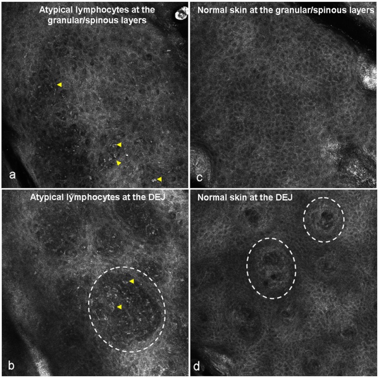
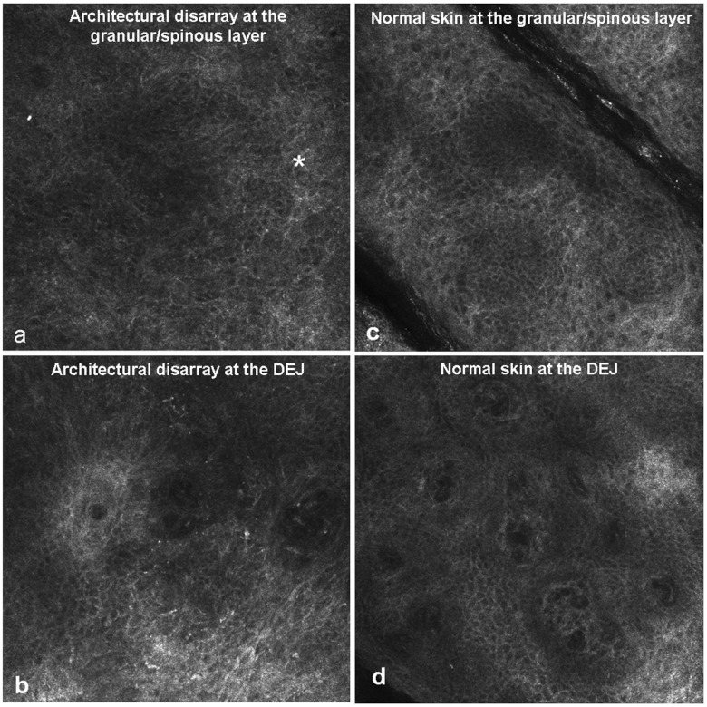
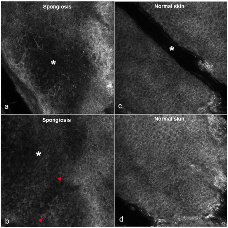
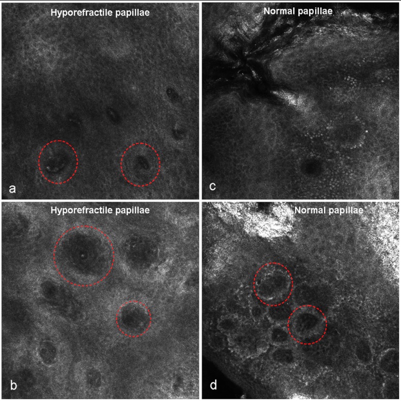
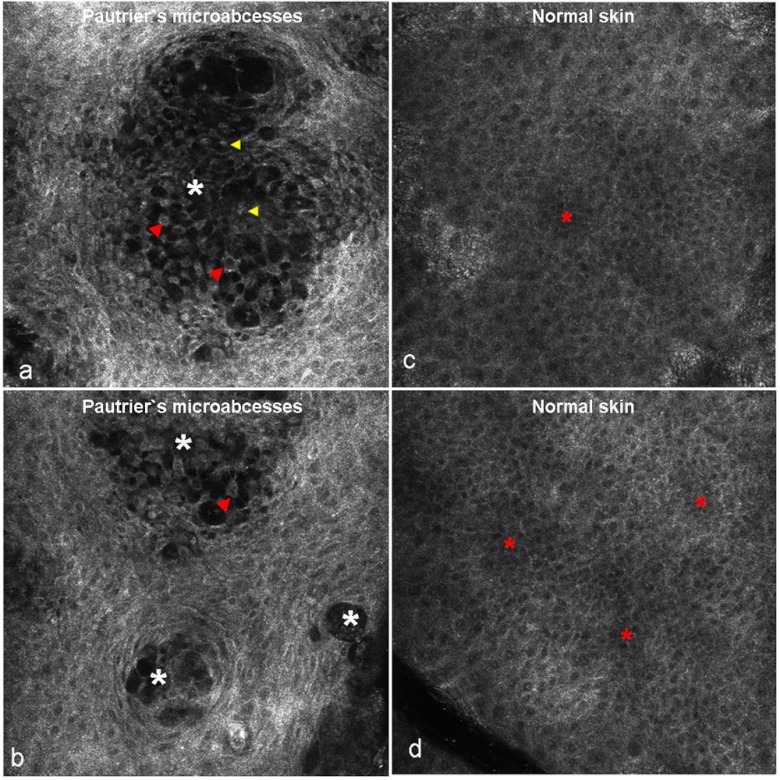
Reflectance confocal microscopy (RCM) represents a noninvasive imaging technique that has previously been used for characterization of mycosis fungoides (MF) in a pilot study. We aimed to test the applicability of RCM for diagnosis and differential diagnosis of MF in a clinical study. A total of 39 test sites of 15 patients with a biopsy-proven diagnosis of either MF, parapsoriasis, Sézary syndrome, or lymphomatoid papulosis were analyzed for presence and absence of RCM features of MF. Cochran and Chi(2) analysis were applied to test the concordance between investigators and the distribution of RCM features, respectively. For selected parameters, the Cochran analysis showed good concordance between investigators. Inter-observer reproducibility was highest for junctional atypical lymphocytes, architectural disarray, and spongiosis. Similarly, Chi(2) analysis demonstrated that selected features were present at particularly high frequency in individual skin diseases, with values ranging from 73% to 100% of all examined cases.
Figures
Similar articles
-
Reflectance confocal microscopy for the characterization of mycosis fungoides and correlation with histology: a pilot study.Skin Res Technol. 2013 Aug;19(3):352-5. doi: 10.1111/srt.12049. Epub 2013 Apr 18. Skin Res Technol. 2013. PMID: 23594100
-
Systematic review and proposal of an in vivo reflectance confocal microscopy assessment tool for cutaneous lymphoma.J Cutan Pathol. 2020 Mar;47(3):295-304. doi: 10.1111/cup.13598. Epub 2019 Nov 6. J Cutan Pathol. 2020. PMID: 31618473
-
In vivo reflectance confocal microscopy of mycosis fungoides: A preliminary study.J Am Acad Dermatol. 2007 Sep;57(3):435-41. doi: 10.1016/j.jaad.2007.02.026. Epub 2007 Apr 16. J Am Acad Dermatol. 2007. PMID: 17433849
-
Concordance between in vivo reflectance confocal microscopy and optical histology of lymphomatoid papulosis.Skin Res Technol. 2013 Aug;19(3):308-13. doi: 10.1111/srt.12046. Epub 2013 Feb 26. Skin Res Technol. 2013. PMID: 23441678
-
Primary cutaneous T-cell lymphoma (mycosis fungoides and Sézary syndrome): part I. Diagnosis: clinical and histopathologic features and new molecular and biologic markers.J Am Acad Dermatol. 2014 Feb;70(2):205.e1-16; quiz 221-2. doi: 10.1016/j.jaad.2013.07.049. J Am Acad Dermatol. 2014. PMID: 24438969 Review.
Cited by
-
Non-Invasive Imaging Including Line-Field Confocal Optical Coherence Tomography (LC-OCT) for Diagnosis of Cutaneous Lymphomas.Cancers (Basel). 2024 Oct 25;16(21):3608. doi: 10.3390/cancers16213608. Cancers (Basel). 2024. PMID: 39518050 Free PMC article.
-
Reflectance confocal microscopy features of mycosis fungoides and Sézary syndrome: correlation with histopathologic and T-cell receptor rearrangement studies.J Cutan Pathol. 2016 Jun;43(6):505-15. doi: 10.1111/cup.12708. Epub 2016 Apr 8. J Cutan Pathol. 2016. PMID: 26969149 Free PMC article. Clinical Trial.
-
Non-invasive Skin Imaging in Cutaneous Lymphomas.Am J Clin Dermatol. 2024 Jan;25(1):79-89. doi: 10.1007/s40257-023-00824-1. Epub 2023 Nov 14. Am J Clin Dermatol. 2024. PMID: 37964050 Free PMC article. Review.
-
Cutaneous T-cell Lymphoma.Clin Lab Med. 2017 Sep;37(3):527-546. doi: 10.1016/j.cll.2017.06.006. Clin Lab Med. 2017. PMID: 28802499 Free PMC article. Review.
-
Non-invasive techniques in the clinical diagnosis of cutaneous lymphomas.Dermatol Reports. 2023 Oct 16;16(Suppl 2):9814. doi: 10.4081/dr.2023.9814. eCollection 2024 May 7. Dermatol Reports. 2023. PMID: 39295885 Free PMC article. Review.
References
-
- Humme D., Lukowsky A., Sterry W., “Diagnostic tools in mycosis fungoides,” G. Ital. Dermatol. Venereol. 145(3), 375–384 (2005).GIDVDZ - PubMed
