. 2012 Jan;17(1):016015.
doi: 10.1117/1.JBO.17.1.016015.

Multifunctional nanoprobe to enhance the utility of optical based imaging techniques

Affiliations

Multifunctional nanoprobe to enhance the utility of optical based imaging techniques

Yeongri Jung et al. J Biomed Opt. 2012 Jan.

Abstract

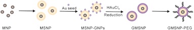
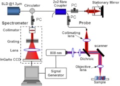
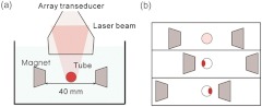
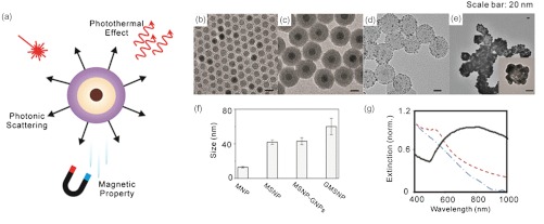
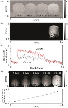
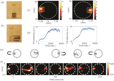
Several imaging modalities such as optical coherence tomography, photothermal, photoacoustic and magnetic resonance imaging, are sensitive to different physical properties (i.e. scattering, absorption and magnetic) that can provide contrast within biological tissues. Usually exogenous agents are designed with specific properties to provide contrast for these imaging methods. In nano-biotechnology there is a need to combine several of these properties into a single contrast agent. This multifunctional contrast agent can then be used by various imaging techniques simultaneously or can be used to develop new imaging modalities. We reported and characterized a multifunctional nanoparticle, made from gold nanoshells, which exhibits scattering, photothermal, photoacoustic, and magnetic properties.


Figures

Fig. 1
Fig. 1
Schematic illustration of GMSNP synthesis.
Fig. 2
Fig. 2
Schematic illustration of photothermal OCT for imaging GMSNP.
Fig. 3
Fig. 3
(a) Experimental setup of the mmPA imaging system. (b) Translation of the magnet system for dynamic magnetic manipulation of GMSNP.
Fig. 4
Fig. 4
(a) Illustration of the multifunctional properties of GMSNP as a contrast agent for optical imaging techniques. Transmission electron microscopy images of (b) MNP, (c) MSNP, (d) MSNP-GNRs (e) GMSNP. (f) The size distribution of each nanoparticle. (g) UV-Vis-NIR extinction of MNP, MSNP and GMSNP. The characteristic absorbance of GMSNP is red-shifted due to the outer gold shell.
Fig. 5
Fig. 5
(a) Conventional scattering and (b) photothermal OCT images of control, MSNP, MSNP-GNPs and GMSNP at a concentration of 5.0 nM. (c) depth-dependent OCT intensity decay of control and 5.0 nM GMSNP. (d) At different concentrations of GMSNP, photothermal images show distinguishable contrast, even though the concentration increases over a small range (left: 0.5 nM, middle: 1.5 nM, Right: 2.5 nM). (e) Plot of photothermal signal strength at the center of each phantom in (d).
Fig. 6
Fig. 6
Photograph of GMSNP (a) without and (b) with a magnetic field applied. (c) PA images of accumulated GMSNP when left (left)/right (right) magnet is closer to the tube. (d) Mean PA intensity versus time of accumulated GMSNP obtained by computing the mean value within the left and right rectangular region in (c). The rectangular region is 0.5×0.5  mm2 (vertical x horizontal). (e), (f) Dynamic manipulation of accumulated GMSNP by changing the positions of the magnets left and right.

Similar articles

Cited by

References

    1. Goldberg B. B., Liu J.B, Forsberg F. “Ultrasound contrast agents: a review,” Ultrasound Med. Biol. 20(4), 319–333 (1994).USMBA310.1016/0301-5629(94)90001-9 - DOI - PubMed
    1. Runge V., et al. , “Paramagnetic agents for contrast-enhanced NMR imaging: a review,” Am. J. Roentgenol. 141(6), 1209–1215 (1983).AJROAM - PubMed
    1. Brigger I., Dubernet C., Couvreur P., “Nanoparticles in cancer therapy and diagnosis,” Adv. Drug Deliv. Rev. 54(5), 631–651 (2002).ADDREP10.1016/S0169-409X(02)00044-3 - DOI - PubMed
    1. Michalet X., et al. , “Quantum dots for live cells, in vivo imaging, and diagnostics,” Science 307(5709), 538–544 (2005).SCIEAS10.1126/science.1104274 - DOI - PMC - PubMed
    1. Zagaynova E. V., et al. , “Contrasting properties of gold nanoparticles for optical coherence tomography: phantom, in vivo studies and Monte Carlo simulation,” Phys. Med. Biol. 53(18), 4995–5009 (2008).PHMBA710.1088/0031-9155/53/18/010 - DOI - PubMed

Publication types