. 2012 May 1;29(7):1455-68.
doi: 10.1089/neu.2011.2207. Epub 2012 Apr 10.

Use-dependent dendritic regrowth is limited after unilateral controlled cortical impact to the forelimb sensorimotor cortex

Affiliations

Use-dependent dendritic regrowth is limited after unilateral controlled cortical impact to the forelimb sensorimotor cortex

Theresa A Jones et al. J Neurotrauma. .

Abstract

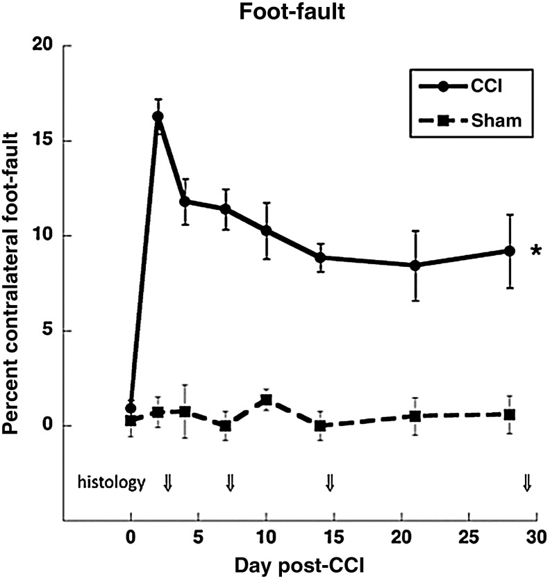
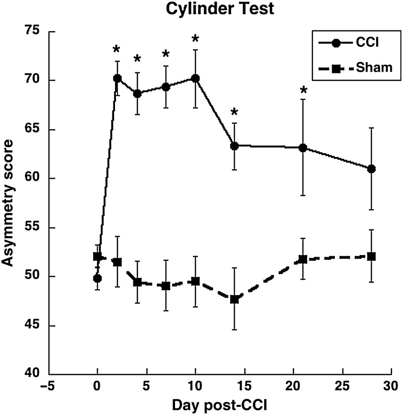
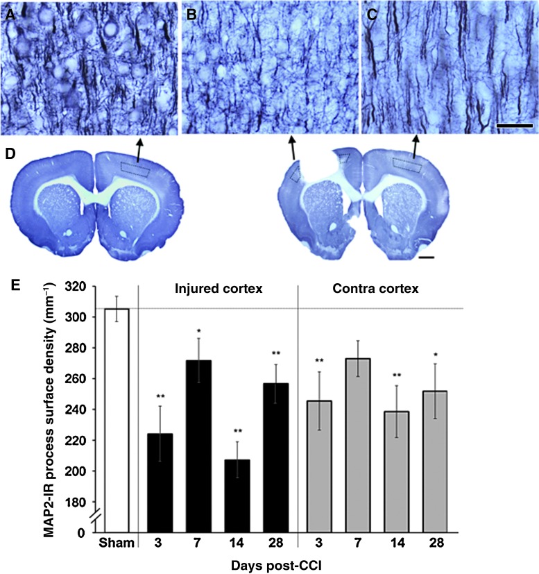
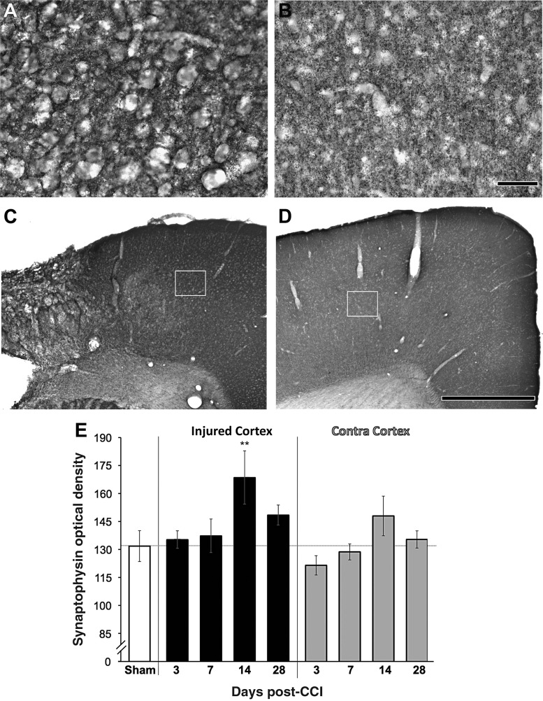
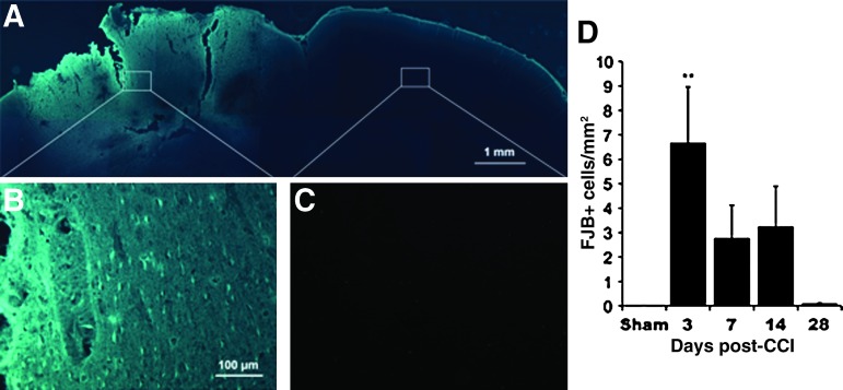
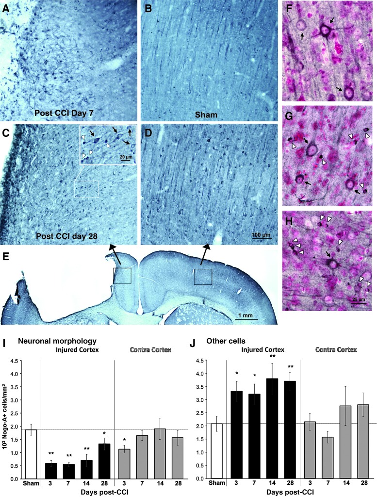
Compensatory neural plasticity occurs in both hemispheres following unilateral cortical damage incurred by seizures, stroke, and focal lesions. Plasticity is thought to play a role in recovery of function, and is important for the utility of rehabilitation strategies. Such effects have not been well described in models of traumatic brain injury (TBI). We examined changes in immunoreactivity for neural structural and plasticity-relevant proteins in the area surrounding a controlled cortical impact (CCI) to the forelimb sensorimotor cortex (FL-SMC), and in the contralateral homotopic cortex over time (3-28 days). CCI resulted in considerable motor deficits in the forelimb contralateral to injury, and increased reliance on the ipsilateral forelimb. The density of dendritic processes, visualized with immunostaining for microtubule-associated protein-2 (MAP-2), were bilaterally decreased at all time points. Synaptophysin (SYN) immunoreactivity increased transiently in the injured hemisphere, but this reflected an atypical labeling pattern, and it was unchanged in the contralateral hemisphere compared to uninjured controls. The lack of compensatory neuronal structural plasticity in the contralateral homotopic cortex, despite behavioral asymmetries, is in contrast to previous findings in stroke models. In the cortex surrounding the injury (but not the contralateral cortex), decreases in dendrites were accompanied by neurodegeneration, as indicated by Fluoro-Jade B (FJB) staining, and increased expression of the growth-inhibitory protein Nogo-A. These studies indicate that, following unilateral CCI, the cortex undergoes neuronal structural degradation in both hemispheres out to 28 days post-injury, which may be indicative of compromised compensatory plasticity. This is likely to be an important consideration in designing therapeutic strategies aimed at enhancing plasticity following TBI.


Conflict of interest statement

No competing financial interests exist.

Figures

FIG. 1.
FIG. 1.
Foot-fault test. All injured animals showed a deficit in forelimb coordination that recovered over time compared to sham animals (*p<0.05 at all time points). Arrows indicate times when animals were removed for histological end-points. Numbers of controlled cortical impact (CCI) animals: days 0–2=41, days 4–7=31, days 10–14=21, days 21 and 28=9 and 10. Numbers of sham animals: days 0–2=14, all other days=8.
FIG. 2.
FIG. 2.
Cylinder test. All injured animals showed a preferential reliance on the non-impaired forelimb for postural support as measured by the asymmetry score: [{(ipsilateral use + ½ bilateral use)/total use} * 100]. Numbers of controlled cortical impact (CCI) animals: days 0 and 2=41 and 39, days 4 and 7=26 and 30, days 10 and 14=21 and 20, days 21 and 28=9 and 10. Numbers of sham animals: days 0 and 2=14, 8 for all remaining time points (*p<0.05 compared to sham animals).
FIG. 3.
FIG. 3.
The density of dendritic arbors (microtubule-associated protein-2, MAP-2) in layer V (AC) were decreased surrounding the injury (B), and in the contralateral homotopic cortex (C), compared to sham animals (A; scale bar=50 μm). Layer V sampling regions are outlined in the coronal sections in (D; scale bar=1 mm). (E) These decreases were seen at all time points following controlled cortical impact (CCI; *p<0.05, **p<0.005 versus sham animals in planned comparisons; at day 7 in the contralateral [Contra] cortex, p=0.08 versus sham animals). Color image is available online at www.liebertonline.com/neu
FIG. 4.
FIG. 4.
Synaptophysin (SYN) immunolabeling in cortex near a controlled cortical impact (CCI; A), and in the contralateral cortex (B; scale bar=50 μm). The white boxes in C and D delineate the regions shown in the higher-magnification images seen in A and B, respectively (scale bar=1 mm). (E) In the injured cortex, there was a transient increase in the optical density of SYN on day 14. No significant changes were found in the contralateral (Contra) cortex (**p=0.004 versus sham animals).
FIG. 5.
FIG. 5.
Fluoro-Jade B (FJB) labeling for degenerating neurons. (A–C) Representative images from 3 days post-CCI. (A) Photomontage of the dorsal portion of a coronal section overexposed to reveal cortical outlines. White boxes outline areas shown in the higher-magnification images seen in B (injured hemisphere) and C (contralateral cortex). (D) Quantification of the density of FJB-labeled neurons. Significant increases were found in the injured cortex at 3 days post-CCI (**p<0.005 versus shams; CCI, controlled cortical impact). Color image is available online at www.liebertonline.com/neu
FIG. 6.
FIG. 6.
Nogo-A labeled cortical samples from 7 days after controlled cortical impact (CCI, A), sham surgery (B), 28 days post-CCI (C), and contralateral cortex 28 days after CCI (D). The inset in C shows examples of cells categorized as having clear neuronal morphology (black arrows) or not (white arrowheads). (E) Lower-magnification view of the sample regions (black boxes) shown in C and D. Counterstaining with pyronin-Y (a Nissl stain) confirmed that cells with neuronal morphology had nuclei characteristic of neurons (F-H). Examples are from the contralateral cortex 28 days after CCI (F), the cortex lateral to the contusion at the same time point (G), and the cortex medial to the contusion on day 7 (H). In the injured hemisphere (I), there was a reduction in the density of Nogo-A-positive cells with obvious neuronal morphology, and an increase in other Nogo-A-positive cells, at all time points. In the contralateral (Contra) cortex (J), significant changes were only found at day 3 post-CCI, and only in Nogo-A-positive cells with neuronal morphology (*p<0.05, *p<0.005 versus sham animals). Color image is available online at www.liebertonline.com/neu

Similar articles

Cited by

References

    1. Adkins D.L. Campos P. Quach D. Borromeo M. Schallert K. Jones T.A. Epidural cortical stimulation enhances motor function after sensorimotor cortical infarcts in rats. Exp. Neurol. 2006;200:356–370. - PubMed
    1. Adkins D.L. Voorhies A.C. Jones T.A. Behavioral and neuroplastic effects of focal endothelin-1 induced sensorimotor cortex lesions. Neuroscience. 2004;128:473–486. - PubMed
    1. Allred R.P. Jones T.A. Experience—a double edged sword for restorative neural plasticity after brain damage. Future Neurol. 2008;3:189–198. - PMC - PubMed
    1. Ansari M.A. Roberts K.N. Scheff S.W. A time course of contusion-induced oxidative stress and synaptic proteins in cortex in a rat model of TBI. J. Neurotrauma. 2008b;25:513–526. - PubMed
    1. Ansari M.A. Roberts K.N. Scheff S.W. Oxidative stress and modification of synaptic proteins in hippocampus after traumatic brain injury. Free Radic. Biol. Med. 2008a;45:443–452. - PMC - PubMed

Publication types

LinkOut - more resources