. 2012 May;46(2):393-401.
doi: 10.1016/j.nbd.2012.02.003. Epub 2012 Feb 14.

JNK2 and JNK3 are major regulators of axonal injury-induced retinal ganglion cell death

Affiliations

JNK2 and JNK3 are major regulators of axonal injury-induced retinal ganglion cell death

Kimberly A Fernandes et al. Neurobiol Dis. 2012 May.

Abstract

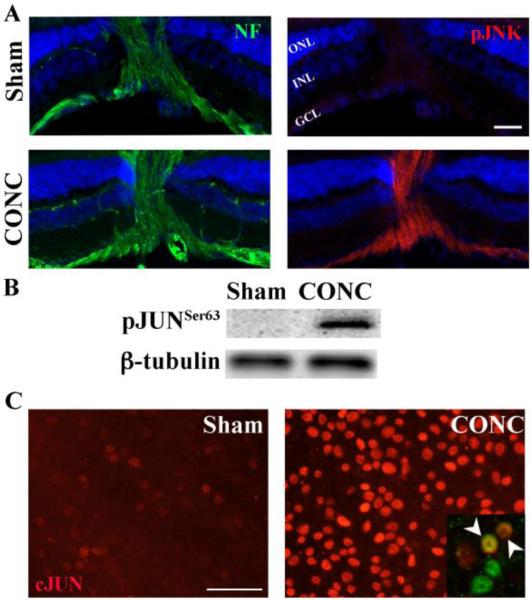
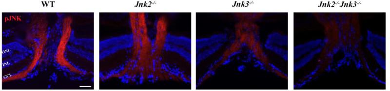
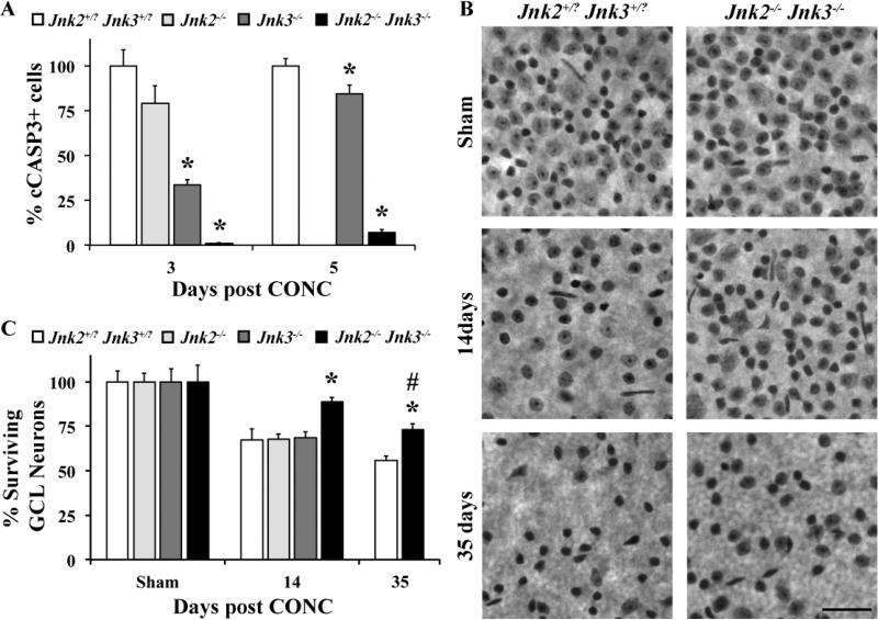
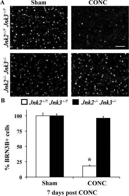
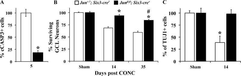
Glaucoma is a neurodegenerative disease characterized by the apoptotic death of retinal ganglion cells (RGCs). The primary insult to RGCs in glaucoma is thought to occur to their axons as they exit the eye in the optic nerve head. However, pathological signaling pathways that exert central roles in triggering RGC death following axonal injury remain unidentified. It is likely that the first changes to occur following axonal injury are signal relay events that transduce the injury signal from the axon to the cell body. Here we focus on the c-Jun N-terminal kinase (JNK1-3) family, a signaling pathway implicated in axonal injury signaling and neurodegenerative apoptosis, and likely to function as a central node in axonal injury-induced RGC death. We show that JNK signaling is activated immediately after axonal injury in RGC axons at the site of injury. Following its early activation, sustained JNK signaling is observed in axonally-injured RGCs in the form of JUN phosphorylation and upregulation. Using mice lacking specific Jnk isoforms, we show that Jnk2 and Jnk3 are the isoforms activated in injured axons. Combined deficiency of Jnk2 and Jnk3 provides robust long-term protection against axonal injury-induced RGC death and prevents downregulation of the RGC marker, BRN3B, and phosphorylation of JUN. Finally, using Jun deficient mice, we show that JUN-dependent pathways are important for axonal injury-induced RGC death. Together these data demonstrate that JNK signaling is the major early pathway triggering RGC death after axonal injury and may directly link axon injury to transcriptional activity that controls RGC death.


Figures

Figure 1
Figure 1. JNK signaling is activated in RGCs in vivo following axonal injury
A, Phosphorylated JNK (pJNK; Red) is not present in the retina or optic nerve head (area where retinal axons, labeled with neurofilament (NF, green) transverse the retina) in the uninjured sham animals. However, 1hr after controlled optic nerve crush injury (CONC), pJNK is in RGC axons (neurofilament positive). B, Western blot analysis shows that phosphorylated JUN (pJUN) is not present in uninjured retinas but is present after CONC (3 days after injury, representative blot, n = 3). C, Immunostaining of retina flatmounts for JUN (red) confirms the upregulation seen by western blot analysis and shows JUN staining in RGCs (Right inset, BRN3B+ cells, green; arrow heads indicate colocalization of BRN3B and JUN). Blue staining in A is DAPI; Scale bar, 50um. ONL, Outer Nuclear Layer; INL, Inner Nuclear Layer; GCL, Ganglion Cell Layer.
Figure 2
Figure 2. Activation of JNK in RGC axons following CONC is attenuated in Jnk3-/- and Jnk2-/-Jnk3-/- retinas
Longitudinal retinal sections through the optic nerve head 1hr following controlled optic nerve crush injury (CONC), immunostained for phosphorylated JNK and the nuclear marker DAPI (blue). Robust activation of JNK is observed in RGC axons of wild-type and Jnk2-/- mice. In contrast, deficiency of Jnk3 or Jnk2&3 dramatically attenuates activation of JNK in RGC axons. Representative images, n = 3. Scale bar, 50um. ONL, Outer Nuclear Layer; INL, Inner Nuclear Layer; GCL, Ganglion Cell Layer.
Figure 3
Figure 3. Deficiency of Jnk2&3 protects RGCs from CONC-induced death
A, At both 3 and 5 days following controlled optic nerve crush (CONC), the percent of dying cells (cleaved caspase 3 positive, cCASP3+) was significantly reduced in the Jnk3-/- and Jnk2-/-Jnk3-/- mice in comparison to the Jnk2+/?Jnk3+/? mice (n ≥ 4 for each genotype and time point; *, P < 0.05, comparing mutant to wildtype mice at same time point). B,C Counts of Nissl stained neurons in retinal flat mounts (RGC side up; representative images shown in B) shows that combined, but not single deficiency of Jnk2 and Jnk3 provides long term protection of RGCs out to at least 35 days (n ≥ 5 for all genotypes and time points). Though, by 35 days it is clear that RGCs are dying in the large numbers in Jnk2-/-Jnk3-/- mice (# P < 0.05, comparing 35 day double mutant mice to Sham of same genotype). Note, only RGCs die after CONC and 40-60% of RGC layer neurons are amacrine cells (Jeon et al., 1998; Li et al., 1999; Li et al., 2007; Quigley et al., 2011); thus, a loss of half of RGC layer neurons is approximately complete RGC loss. Scale bar, 50um.
Figure 4
Figure 4. The RGC-specific marker, BRN3B is not downregulated in Jnk2-/-Jnk3-/- mice
A, Representative images showing BRN3B immunolabeled cells in the GCL of Jnk2+/?Jnk3+/? or Jnk2-/-Jnk3-/- mice 7 days following sham treatment or controlled optic nerve crush (CONC). B,The number of BRN3B positive cells in the Jnk2+/?Jnk3+/? retinas was significantly reduced at 7 days post CONC, reflecting both RGC death and/or BRN3B downregulation. In contrast, in the Jnk2-/-Jnk3-/- mice, the number of BRN3B positive cells at 7 days post CONC was indistinguishable from the number of BRN3B positive cells in sham-treated eyes confirming the protection provided by Jnk2/3 deficiency. n = 3 for each genotype; *, P < 0.001; scale bar, 50um.
Figure 5
Figure 5. Phosphorylation of JUN is attenuated in the Jnk3-/- and Jnk2-/-Jnk3-/- nulls
A, Representative western blot showing the expression of pJUNSer63 3 days post CONC (Ser63 is the residue that is phosphorylated by JNK, (Pulverer et al., 1991). B, Quantification by densitometric analyses of 4 different blots using retinal protein lysates from 4 different eyes shows that the relative amounts of pJUNSer63 (normalized to β-tubulin) is significantly reduced in the Jnk2-/-Jnk3-/- retinas compared to controls. *, P < 0.001.
Figure 6
Figure 6. Deficiency of JUN protects RGCs from CONC-induced death
A, Deletion of Jun in the retina (Junfl/fl; Six3-cre+) significantly reduced (*, P < 0.001) the number of dying cells (cCASP3+) 5 days after CONC compared to Jun+/?; Six3-cre- mice. B, Nissl counts of the number of surviving GCL neurons showed that Jun deficiency protected from axonal-injury induced death at 14 days following CONC (n ≥ 5 per group; *, P < 0.001). Counts of surviving GCL neurons in Junfl/fl; Six3-cre+ mice 35 days following CONC indicate both a increased survival compared to Jun+/?; Six3-cre? mice (*, P = 0.001) and a significant loss compared to the uninjured Junfl/fl; Six3-cre+ mice (#, P = 0.008). C, Counts using the RGC marker TUJ1 at 14 days after injury confirmed both the significant loss of RGCs in the Jun+/?; Six3-cre- mice (*, P < 0.001) and the prevention of RGC death in Junfl/fl; Six3-cre+ retinas.

References

    1. Abe N, Cavalli V. Nerve injury signaling. Current opinion in neurobiology. 2008;18:276–83. - PMC - PubMed
    1. Anderson DR, Hendrickson A. Effect of intraocular pressure on rapid axoplasmic transport in monkey optic nerve. Invest Ophthalmol. 1974;13:771–83. - PubMed
    1. Angel P, et al. The jun proto-oncogene is positively autoregulated by its product, Jun/AP-1. Cell. 1988;55:875–85. - PubMed
    1. Badea TC, et al. Distinct roles of transcription factors brn3a and brn3b in controlling the development, morphology, and function of retinal ganglion cells. Neuron. 2009;61:852–64. - PMC - PubMed
    1. Behrens A, et al. Impaired postnatal hepatocyte proliferation and liver regeneration in mice lacking c-jun in the liver. The EMBO journal. 2002;21:1782–90. - PMC - PubMed

Publication types

MeSH terms

Substances