. 2012 Apr;158(4):1916-32.
doi: 10.1104/pp.111.192021. Epub 2012 Feb 21.

Prunasin hydrolases during fruit development in sweet and bitter almonds

Affiliations

Prunasin hydrolases during fruit development in sweet and bitter almonds

Raquel Sánchez-Pérez et al. Plant Physiol. 2012 Apr.

Abstract

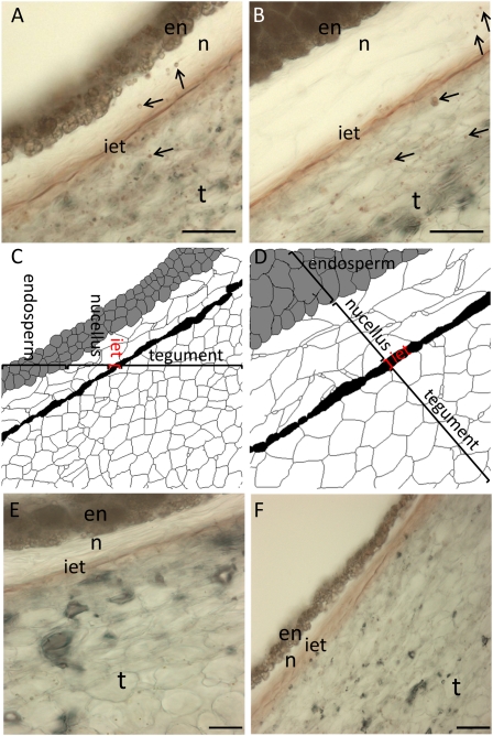
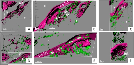
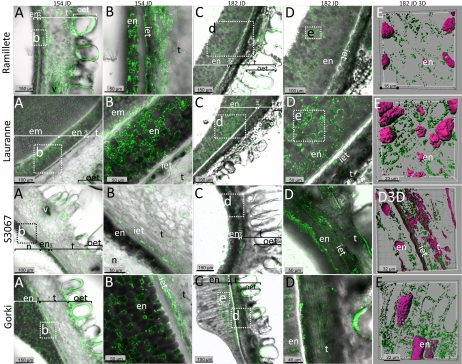
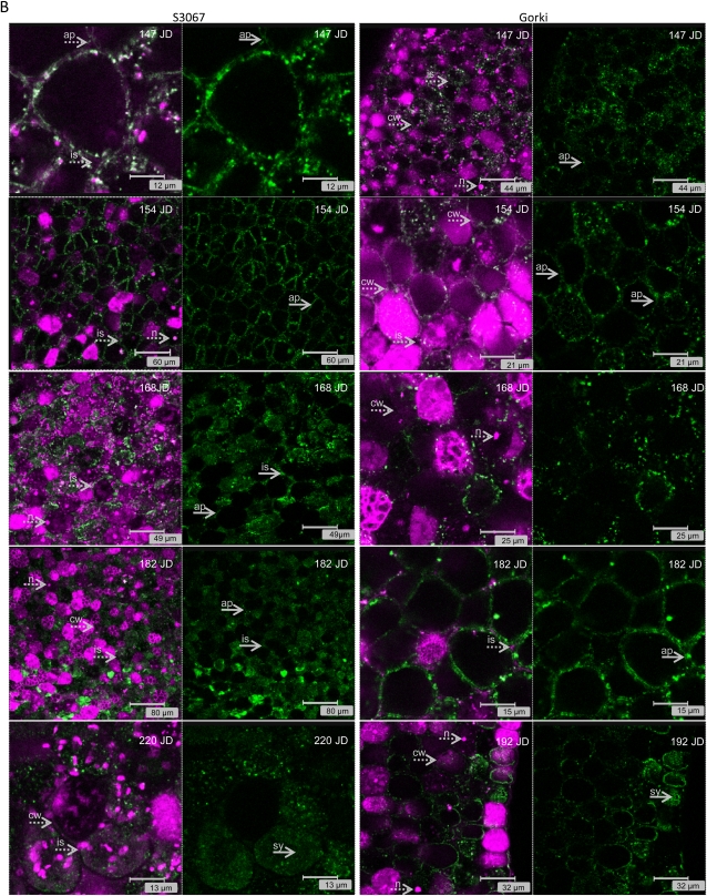
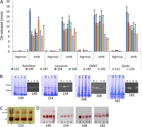
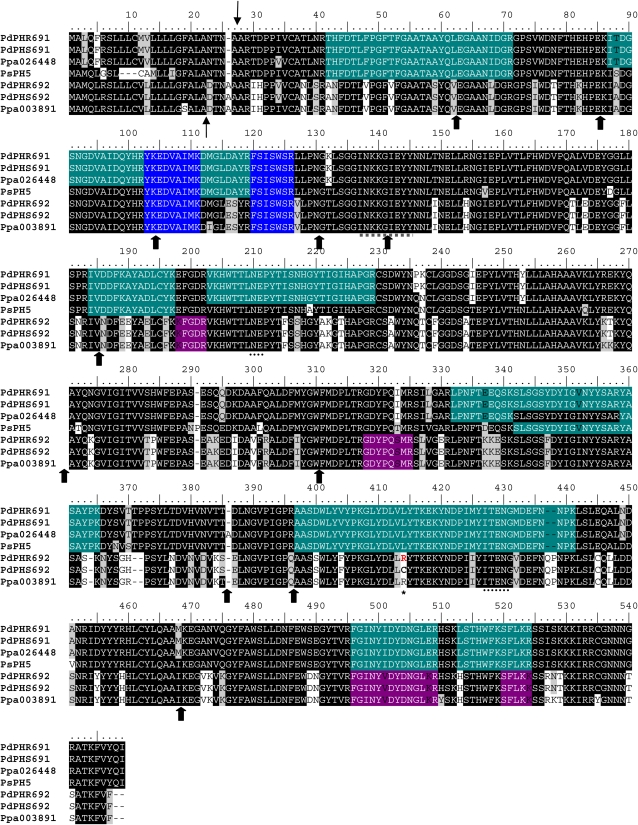
Amygdalin is a cyanogenic diglucoside and constitutes the bitter component in bitter almond (Prunus dulcis). Amygdalin concentration increases in the course of fruit formation. The monoglucoside prunasin is the precursor of amygdalin. Prunasin may be degraded to hydrogen cyanide, glucose, and benzaldehyde by the action of the β-glucosidase prunasin hydrolase (PH) and mandelonitirile lyase or be glucosylated to form amygdalin. The tissue and cellular localization of PHs was determined during fruit development in two sweet and two bitter almond cultivars using a specific antibody toward PHs. Confocal studies on sections of tegument, nucellus, endosperm, and embryo showed that the localization of the PH proteins is dependent on the stage of fruit development, shifting between apoplast and symplast in opposite patterns in sweet and bitter cultivars. Two different PH genes, Ph691 and Ph692, have been identified in a sweet and a bitter almond cultivar. Both cDNAs are 86% identical on the nucleotide level, and their encoded proteins are 79% identical to each other. In addition, Ph691 and Ph692 display 92% and 86% nucleotide identity to Ph1 from black cherry (Prunus serotina). Both proteins were predicted to contain an amino-terminal signal peptide, with the size of 26 amino acid residues for PH691 and 22 residues for PH692. The PH activity and the localization of the respective proteins in vivo differ between cultivars. This implies that there might be different concentrations of prunasin available in the seed for amygdalin synthesis and that these differences may determine whether the mature almond develops into bitter or sweet.


Figures

Figure 1.
Figure 1.
Seeds from two almond cultivars, sweet (Lauranne; A and C) and bitter (S3067; B and D), at 154 JD were cross-sectioned to monitor the distribution of PH activity using the sugar-reducing assay after incubation with prunasin. A and B, PH was detected in the tegument (t), inner epidermis of the tegument (iet), nucellus (n), and endosperm (en), as observed by reddish vesicles indicated with arrows. C and D, Histological diagrams, defining the different type of tissues analyzed in the cross-sections. E and F, Background staining in the absence of added substrate in Lauranne (E) and S3067 (F). Bars = 50 μm. [See online article for color version of this figure.]
Figure 2.
Figure 2.
Seeds from two almond cultivars, sweet (Lauranne; A–C) and bitter (S3067; D–F), at 154 JD were cross-sectioned to perform immunolocalization studies using a polyclonal antibody against the sequence INKKGIEYY present in the PHs from almond (green vesicles). PHs were detected in the tegument (t), inner epidermis of the tegument (iet), nucellus (n), and endosperm (en). Magenta color represents the cell wall (cw) and the DNA/RNA structures stained with propidium iodide in the confocal studies. Arrows indicate the PH vesicles in the cells belonging to the inner epidermis of the tegument. A and D show three-dimensional presentations of the z stack, whereas B, C, E, and F present magnifications of the cells in the inner tegument with the same view and different view angles. A, B, D, and E show views of open cells: in the sweet Lauranne cultivar (A and B), the cell wall of the inner epidermis of the tegument remains devoid of PH vesicles, whereas in the bitter S3067 cultivar (D and E), PH vesicles are observed within the cell wall. [See online article for color version of this figure.]
Figure 3.
Figure 3.
Immunolocalization studies of prunasin localization in four different almond cultivars in the course of fruit development. The cultivars Ramillete (sweet), Lauranne (sweet), S3067 (bitter), and Gorki (bitter) were analyzed for PH content at two different time points during mid season (154 and 182 JD) of fruit development using a polyclonal antibody against the sequence INKKGIEYY present in the PHs (green color) from almond. The first four columns represent overlay images of PH detected by the specific antibody (green) and translucent light micrographs to visualize cell walls (gray). The marked area in a panel represents the area shown at higher magnification in the panel to the right in the same row, except for Gorki, where the source of the images in columns d and e are derived from the marked segments in column c. The last column represents three-dimensional images obtained using sections containing mainly endosperm cells stained with propidium iodide in magenta color and green vesicles that represent the PHs. Abbreviations are as follows: endosperm (en), inner epidermis of the tegument (iet), tegument (t), sometimes with vascular bundles (v), and outer epidermis of the tegument (out). In a small number of the sections, the embryo (em) or the nucellus (n) remained attached to the rest of the tissue. In the later stage shown (182 JD), the tegument cells have collapsed. [See online article for color version of this figure.]
Figure 4.
Figure 4.
(Legend appears on following page.) Figure 4. A, Immunolocalization studies of PHs (green color) in the embryo of two sweet genotypes (Ramillete and Lauranne) at different time points during fruit development (Ramillete, 147, 154, 168, 182, and 220 JD; Lauranne, 147, 154, 168, 182, and 192 JD) using a polyclonal antibody against the sequence INKKGIEYY present in the PHs from almond. The presence of DNA/RNA structures (i.e. nucleus [n]) and cell wall (cw) is visualized by propidium iodide staining (magenta color) and indicated by dotted arrows. The localization of the PH changes during fruit development from being mainly in the symplast (sy; i.e. Ramillete at 147, 154, and 168 JD and Lauranne at 147, 154, and 168 JD) to being predominantly found in the apoplast (ap; i.e. Ramillete at 182 and 192 JD and Lauranne at 182 JD), as indicated by solid arrows, filling even the intercellular spaces (is). B, Immunolocalization studies of PHs (green color) in the cotyledon of two bitter genotypes (S3067 and Gorki) at different time points during fruit development (S3067, 147, 154, 168, 182, and 220 JD; Gorki, 147, 154, 168, 182, and 192 JD) using a polyclonal antibody against the sequence INKKGIEYY present in the PHs from almond. The presence of DNA/RNA structures (i.e. nucleus [n]) and cell wall (cw) is visualized by propidium iodide staining (magenta color) and indicated by dotted arrows. The localization of the PH changes during fruit development from being mainly in the apoplast (i.e. S3067 at 147,154, 168, and 182 JD and Gorki at 147, 154, 168, and 182 JD), with occasional presence in the intercellular spaces (is), to being found in the symplast (i.e. S3067 at 220 JD and Gorki at 192 JD). [See online article for color version of this figure.]
Figure 4.
Figure 4.
(Legend appears on following page.) Figure 4. A, Immunolocalization studies of PHs (green color) in the embryo of two sweet genotypes (Ramillete and Lauranne) at different time points during fruit development (Ramillete, 147, 154, 168, 182, and 220 JD; Lauranne, 147, 154, 168, 182, and 192 JD) using a polyclonal antibody against the sequence INKKGIEYY present in the PHs from almond. The presence of DNA/RNA structures (i.e. nucleus [n]) and cell wall (cw) is visualized by propidium iodide staining (magenta color) and indicated by dotted arrows. The localization of the PH changes during fruit development from being mainly in the symplast (sy; i.e. Ramillete at 147, 154, and 168 JD and Lauranne at 147, 154, and 168 JD) to being predominantly found in the apoplast (ap; i.e. Ramillete at 182 and 192 JD and Lauranne at 182 JD), as indicated by solid arrows, filling even the intercellular spaces (is). B, Immunolocalization studies of PHs (green color) in the cotyledon of two bitter genotypes (S3067 and Gorki) at different time points during fruit development (S3067, 147, 154, 168, 182, and 220 JD; Gorki, 147, 154, 168, 182, and 192 JD) using a polyclonal antibody against the sequence INKKGIEYY present in the PHs from almond. The presence of DNA/RNA structures (i.e. nucleus [n]) and cell wall (cw) is visualized by propidium iodide staining (magenta color) and indicated by dotted arrows. The localization of the PH changes during fruit development from being mainly in the apoplast (i.e. S3067 at 147,154, 168, and 182 JD and Gorki at 147, 154, 168, and 182 JD), with occasional presence in the intercellular spaces (is), to being found in the symplast (i.e. S3067 at 220 JD and Gorki at 192 JD). [See online article for color version of this figure.]
Figure 5.
Figure 5.
A, HCN released (nmol) from the tegument and nucellus (teg+nuc) and embryo (emb) in two sweet cultivars (Ramillete [R] and Lauranne [L]) and two bitter cultivars (S3067 [S] and Gorki [G]) following incubation of crude protein extracts with 20 nmol of prunasin at different stages of fruit development (133–220 JD). B, Total protein extracts were analyzed by SDS-PAGE, stained with Coomassie Brilliant Blue, and PHs present were visualized by immunolocalization using a polyclonal antibody against the sequence INKKGIEYY present in the PHs from almond. C, Total β-glucosidase content was assayed using Fast Blue BB salt staining of SDS-PAGE following the application of crude protein extracts of cotyledons of the four different cultivars harvested at 133 JD and revealed the presence of different isoforms of PHs (PH) and the presence of amygdalin hydrolase (AH). D, The same assay was used to monitor the presence of different β-glucosidases during fruit development in the four cultivars. [See online article for color version of this figure.]
Figure 6.
Figure 6.
(Legend appears on following page.) Figure 6. Alignment of different PHs from the P. dulcis (Pd) isoforms 691 and 692 (PHR, Ramillete; PHS, S3067) identified in this study with black cherry isoform 5 (PsPH5) and peach (Ppa) 026448 and 003891. Thin arrows mark the predicted cleavage site of the signal peptide in the almond PHs. Thick arrows represent the splicing sites for the 12 introns for the almond sequences, characteristic for the PHs. ITENG and NEP motifs diagnostic for glycosyl hydrolase family 1 are marked by dotted lines. The peptide used to generate the polyclonal antibody in rabbit is underlined with a dotted gray line. The peptides identified by LC-MS/MS are highlighted in green (for the ones present in PH691), purple (PH692), and blue (present in all the sequences). The only difference between sequences from sweet and bitter cultivars was detected in PdPH692, at position 414 (asterisk). [See online article for color version of this figure.]

Similar articles

Cited by

References

    1. Andréasson E, Bolt Jørgensen L, Höglund A-S, Rask L, Meijer J. (2001) Different myrosinase and idioblast distribution in Arabidopsis and Brassica napus. Plant Physiol 127: 1750–1763 - PMC - PubMed
    1. Arrázola G. (2002) Análisis de glucósidos cianogénicos en variedades de almendro: implicaciones en la mejora genética. PhD thesis. Universidad de Alicante, Alicante, Spain
    1. Ben-Yehoshua S, Conn EE. (1964) Biosynthesis of prunasin, the cyanogenic glucoside of peach. Plant Physiol 39: 331–333 - PMC - PubMed
    1. Conn EE. (1980) Cyanogenic compounds. Annu Rev Plant Physiol 31: 433–451
    1. Dicenta F, Martínez-Gómez P, Grané N, Martín ML, León A, Cánovas JA, Berenguer V. (2002) Relationship between cyanogenic compounds in kernels, leaves, and roots of sweet and bitter kernelled almonds. J Agric Food Chem 50: 2149–2152 - PubMed

Publication types

MeSH terms

LinkOut - more resources