Review
. 2012 Dec 31;327(1-2):61-72.
doi: 10.1016/j.canlet.2012.02.001. Epub 2012 Feb 19.

Pathways for repairing and tolerating the spectrum of oxidative DNA lesions

Affiliations
Review

Pathways for repairing and tolerating the spectrum of oxidative DNA lesions

Brian R Berquist et al. Cancer Lett. .

Abstract

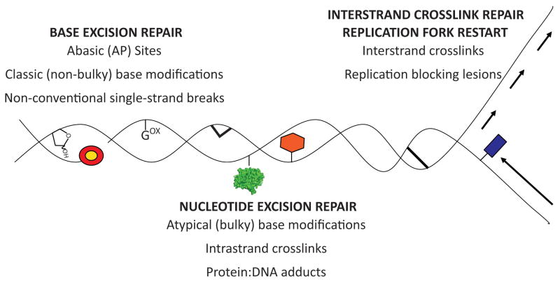
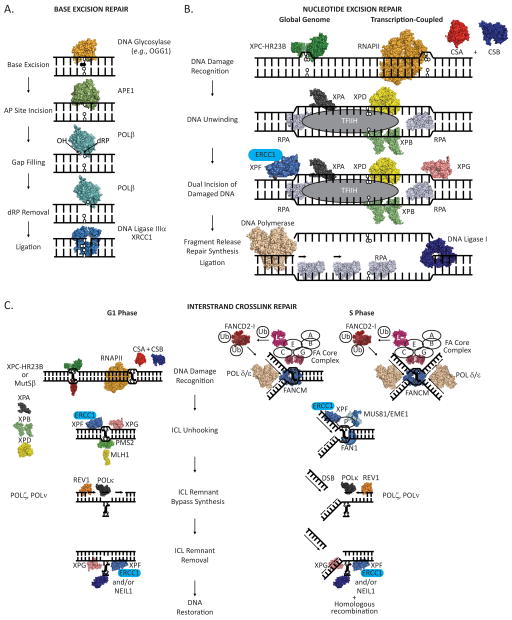
Reactive oxygen species (ROS) arise from both endogenous and exogenous sources. These reactive molecules possess the ability to damage both the DNA nucleobases and the sugar phosphate backbone, leading to a wide spectrum of lesions, including non-bulky (8-oxoguanine and formamidopyrimidine) and bulky (cyclopurine and etheno adducts) base modifications, abasic sites, non-conventional single-strand breaks, protein-DNA adducts, and intra/interstrand DNA crosslinks. Unrepaired oxidative DNA damage can result in bypass mutagenesis during genome copying or gene expression, or blockage of the essential cellular processes of DNA replication or transcription. Such outcomes underlie numerous pathologies, including, but not limited to, carcinogenesis and neurodegeneration, as well as the aging process. Cells have adapted and evolved defense systems against the deleterious effects of ROS, and specifically devote a number of cellular DNA repair and tolerance pathways to combat oxidative DNA damage. Defects in these protective pathways trigger hereditary human diseases that exhibit increased cancer incidence, developmental defects, neurological abnormalities, and/or premature aging. We review herein classic and atypical oxidative DNA lesions, outcomes of encountering these damages during DNA replication and transcription, and the consequences of losing the ability to repair the different forms of oxidative DNA damage. We particularly focus on the hereditary human diseases Xeroderma Pigmentosum, Cockayne Syndrome and Fanconi Anemia, which may involve defects in the efficient repair of oxidative modifications to chromosomal DNA.


Figures

Figure 1
Figure 1
Classic and atypical forms of oxidative DNA damage and associated DNA repair pathway(s). Gox = 8-oxoG; red/yellow circle = 5′ or 3′ terminal blocking group; green protein = protein-DNA adduct; red hexagon = bulky base modification; thick lines = intra- or inter-strand crosslink; blue rectangle = replication blocking lesion.
Figure 2
Figure 2
(A) Major steps in the short-patch base excision repair (BER) pathway. A damaged base is detected and excised from the DNA duplex by a DNA glycosylase, leaving an apurinic/apyrimidinic site that can be bound by APE1 endonuclease. APE1 then incises the DNA phosphodiester backbone 5′ to the abasic site leaving a 3′-hydroxyl (OH) and a 5′-deoxyribose phosphate (dRP). DNA Pol β removes the dRP moiety, via an intrinsic dRP lyase activity, and inserts a nucleotide. The remaining nick in the phosphodiester backbone is sealed through the action of a DNA ligase, leaving intact, undamaged duplex DNA. (B) Major steps in the nucleotide excision repair (NER) pathway. Helix distorting DNA damage (an oxidatively damaged DNA base in this instance) can be recognized by the XPC-HR23B protein complex (global genome-NER) or by an elongating RNA polymerase in concert with the CSA and CSB proteins (transcription-coupled-NER). XPA, RPA, TFIIH (including the XPB and XPD ATPases/helicases) are recruited to the site of the damage, and the DNA surrounding the damage is unwound. XPG and XPF/ERCC1 nucleases are recruited and cleave the DNA phosphodiester backbone on the 3′ and 5′ sides of the damage, respectively, releasing a damage containing DNA section. A DNA polymerase is then enlisted to fill the gap, leaving a nick in the phosphodiester backbone that is sealed by a DNA ligase, producing intact, undamaged duplex DNA. (C) Model of major steps in the repair of interstrand crosslinks (ICLs). In the G1 phase of the cell cycle, an ICL can be recognized by XPC-HR23B protein complex, MutSβ (MSH2-MSH3) protein complex, or by an elongating RNA polymerase in concert with the CSA and CSB proteins. Once the ICL has been recognized, it is unhooked from one strand by the action of the classic NER pathway, which involves the XPG and XPF/ERCC1 nucleases, or potentially through the endonuclease activity of the MLH1/PMS2 protein complex. The ICL remnant is then bypassed through the action of translesion DNA polymerases: REV1 then POLκ, and/or POLζ, POLν, and possibly additional DNA polymerases. At this point, a second round of DNA repair is initiated to remove the covalently linked ICL remnant from the second DNA strand. NER, involving the nuclease actions of XPG and XPF/ERCC1, likely acts to remove a DNA segment containing the physically linked ICL remnant, or the ICL remnant can be removed through the action of the NEIL1 DNA glycosylase. DNA can then be restored to the native duplex form through the activity of DNA polymerase(s), to fill in the gap, and a DNA ligase, to seal the nick in the phosphodiester backbone. During the S phase of a cell cycle, an ICL is detected by stalling of a single replication fork (left) or two converging replication forks (right). FANCM is initially recruited to the stalled replication fork complex and likely acts to regress and or stabilize the fork. The FA core complex, comprised of FANCA, B, C, E, F, G and L, recognizes the FANCM bound/stabilized replication fork and ubiquitinates the FANCD2/I protein complex, with FANCL acting as the E3 ubiquitin ligase. Ubiquitinated FANCD2/I then associates with chromatin. For simplicity, action at a single replication fork is shown further. FANCP coordinates endonucleolytic cleavage of the ICL stalled replication fork by XPF/ERCC1, MUS81/EME1, and FAN1. Cleavage by these nucleases unhooks the ICL from one DNA strand and generates a double strand break (DSB). DNA polymerization past the unhooked ICL remnant by translesion DNA polymerases, REV1 then POLκ, and/or POLζ, POLν, and possibly additional DNA polymerases, ensues. A second round of repair is initiated to remove the unhooked ICL remnant by NER and/or NEIL1. Homologous recombination then is used to reform the replication fork.

References

    1. Imlay JA, Linn S. DNA damage and oxygen radical toxicity. Science. 1988;240:1302–1309. - PubMed
    1. Mannaerts GP, Van Veldhoven PP. Metabolic pathways in mammalian peroxisomes. Biochimie. 1993;75:147–158. - PubMed
    1. Shen Z, Wu W, Hazen SL. Activated leukocytes oxidatively damage DNA, RNA, and the nucleotide pool through halide-dependent formation of hydroxyl radical. Biochemistry. 2000;39:5474–5482. - PubMed
    1. Kovacic P, Wakelin LP. Review: DNA molecular electrostatic potential: novel perspectives for the mechanism of action of anticancer drugs involving electron transfer and oxidative stress. Anticancer Drug Des. 2001;16:175–184. - PubMed
    1. Wood ML, Dizdaroglu M, Gajewski E, Essigmann JM. Mechanistic studies of ionizing radiation and oxidative mutagenesis: genetic effects of a single 8-hydroxyguanine (7-hydro-8-oxoguanine) residue inserted at a unique site in a viral genome. Biochemistry. 1990;29:7024–7032. - PubMed

Publication types

Substances