. 2012 Feb 21:3:687.
doi: 10.1038/ncomms1693.

The dynamic stator stalk of rotary ATPases

Affiliations
Free PMC article

The dynamic stator stalk of rotary ATPases

Alastair G Stewart et al. Nat Commun. .
Free PMC article

Abstract

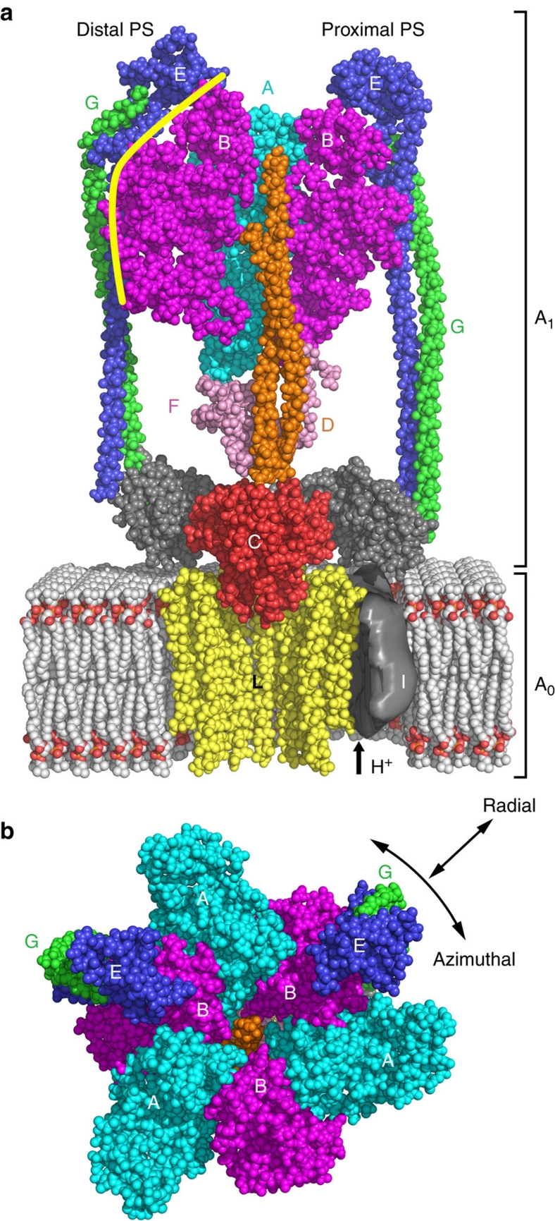
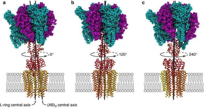
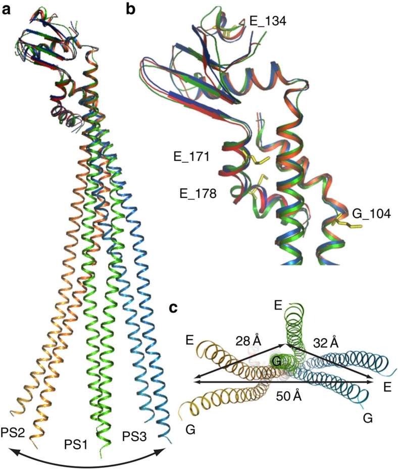
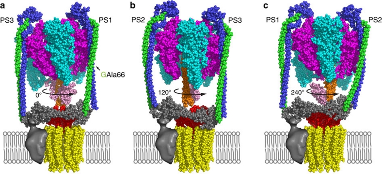
Rotary ATPases couple ATP hydrolysis/synthesis with proton translocation across biological membranes and so are central components of the biological energy conversion machinery. Their peripheral stalks are essential components that counteract torque generated by rotation of the central stalk during ATP synthesis or hydrolysis. Here we present a 2.25-Å resolution crystal structure of the peripheral stalk from Thermus thermophilus A-type ATPase/synthase. We identify bending and twisting motions inherent within the structure that accommodate and complement a radial wobbling of the ATPase headgroup as it progresses through its catalytic cycles, while still retaining azimuthal stiffness necessary to counteract rotation of the central stalk. The conformational freedom of the peripheral stalk is dictated by its unusual right-handed coiled-coil architecture, which is in principle conserved across all rotary ATPases. In context of the intact enzyme, the dynamics of the peripheral stalks provides a potential mechanism for cooperativity between distant parts of rotary ATPases.


Figures

Figure 1
Figure 1. Model of the intact A-ATPase obtained by fitting individual structures into the EM density of the intact T. thermophilus complex.
(a) Side view: subunit A is shown in cyan, B in magenta, C in red, D in orange, F in pink, L in yellow, E in blue, G in green and soluble I in grey. Two A subunits and one B subunit were omitted for clarity. As there is currently no structure for the L-ring available in the protein data bank, we modified the theoretical model of a NMR-derived E. coli c12-ring (pdb entry: 1c17) to give a symmetrical c12-ring. The unknown structure of the transmembrane portion of subunit I is indicated by a cut-out EM density in grey, with the proton path indicated by an arrow. (b) Top view: Explanation of the definitions of 'radial' and 'azimuthal' as used in the text. PDB codes of coordinates used are: 3a5d: T. thermophilus (AB)3DF complex, 1r5z: T. thermophilus subunit C, 3k5b: T. thermophilus EG complex (PS1) , 3rrk: Myotis ruber soluble I and 1c17: E. coli subunit c12-ring model.
Figure 2
Figure 2. The three rotational states of the composite model of the T. thermophilus A-ATPase.
The central rotor (subunits D, F, C and the L-ring) is rotated by 0° (a), 120° (b) and 240° (c) about the central axis through the L-ring (as indicated) relative to the (AB)3 ring. The central axis through the centre of the (AB)3 ring is inclined with respect to the central axis through the L-ring (∼6° tilt).
Figure 3
Figure 3. Comparison of peripheral stalk structures.
PS1, PS2 and PS3 were superposed by their globular domains (using residues 100–188 of subunit E). PS1 is shown in green, PS2 in yellow to red colours and PS3 is shown in blue. (a) View from side, with black arrow representing maximum flexion between PS2 and PS3. The tail flexes primarily in a plane corresponding to a radial direction in the intact A-ATPase. (b) Superposition of peripheral stalk globular heads, with methionines present in PS1 highlighted in yellow and labelled, and (c) view from bottom, with black arrows indicating maximum displacement of helices.
Figure 4
Figure 4. Rotating pseudo-atomic model of the A-ATPase with the three peripheral stalks docked at corresponding positions.
(a) 0°, (b) 120° and (c) 240° rotation of the central stalk relative to the stator subunits showing how the peripheral stalk dynamics complements the shape of the (AB)3 domain during the rotary catalytic cycle. Residue Ala66 of subunit G is highlighted to indicate the transition from hendecad to quindecad repeat in the peripheral stalk. The pdb file for the model shown in (a) is supplied as Supplementary Information.

References

    1. Muench S. P., Trinick J. & Harrison M. A. Structural divergence of the rotary ATPases. Q. Rev. Biophys. 44, 311–356 (2011). - PubMed
    1. Cross R. L. & Muller V. The evolution of A-, F-, and V-type ATP synthases and ATPases: reversals in function and changes in the H+/ATP coupling ratio. FEBS Lett. 576, 1–4 (2004). - PubMed
    1. Yoshida M., Muneyuki E. & Hisabori T. ATP synthase--a marvellous rotary engine of the cell. Nat. Rev. Mol. Cell. Biol. 2, 669–677 (2001). - PubMed
    1. Jefferies K. C., Cipriano D. J. & Forgac M. Function, structure and regulation of the vacuolar (H+)-ATPases. Arch. Biochem. Biophys. 476, 33–42 (2008). - PMC - PubMed
    1. Nakano M. et al.. ATP hydrolysis and synthesis of a rotary motor V-ATPase from Thermus thermophilus. J. Biol. Chem. 283, 20789–20796 (2008). - PMC - PubMed

Publication types

MeSH terms

Associated data