. 2012 May 1;365(1):50-60.
doi: 10.1016/j.ydbio.2012.02.008. Epub 2012 Feb 12.

Neurotrophin-4 regulates the survival of gustatory neurons earlier in development using a different mechanism than brain-derived neurotrophic factor

Affiliations

Neurotrophin-4 regulates the survival of gustatory neurons earlier in development using a different mechanism than brain-derived neurotrophic factor

Ami V Patel et al. Dev Biol. .

Abstract

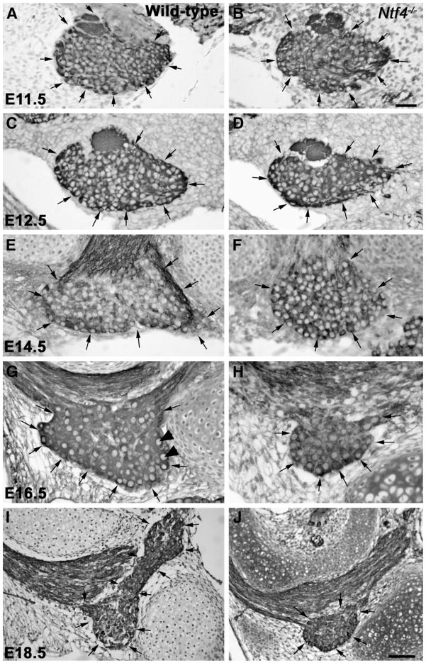
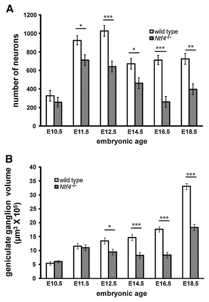
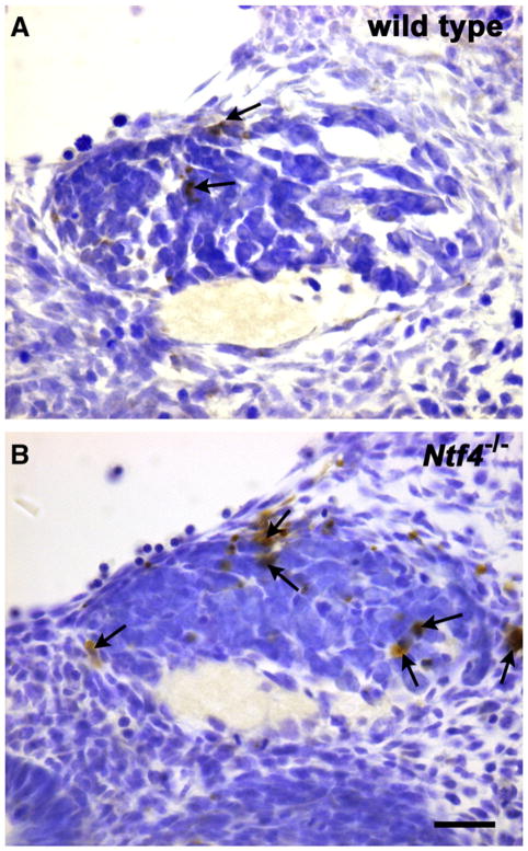
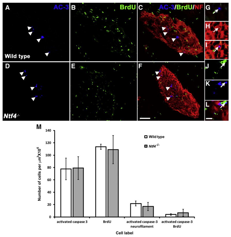
The number of neurons in the geniculate ganglion that are available to innervate taste buds is regulated by neurotrophin-4 (NT-4) and brain-derived neurotrophic factor (BDNF). Our goal for the current study was to examine the timing and mechanism of NT-4-mediated regulation of geniculate neuron number during development. We discovered that NT-4 mutant mice lose 33% of their geniculate neuronal cells between E10.5 and E11.5. By E11.5, geniculate axons have just reached the tongue and do not yet innervate their gustatory targets; thus, NT-4 does not function as a target-derived growth factor. At E11.5, no difference was observed in proliferating cells or the rate at which cells exit the cell cycle between NT-4 mutant and wild type ganglia. Instead, there was an increase in TUNEL-labeling, indicating an increase in cell death in Ntf4(-/-) mice compared with wild types. However, activated caspase-3, which is up-regulated in the absence of BDNF, was not increased. This finding indicates that cell death initiated by NT-4-removal occurs through a different cell death pathway than BDNF-removal. We observed no additional postnatal loss of taste buds or neurons in Ntf4(-/-) mice. Thus, during early embryonic development, NT-4 produced in the ganglion and along the projection pathway inhibits cell death through an activated caspase-3 independent mechanism. Therefore, compared to BDNF, NT-4 plays distinct roles in gustatory development; differences include timing, source of neurotrophin, and mechanism of action.


Figures

Fig. 1
Fig. 1
Anti-TUJ-1-labeled geniculate ganglia (arrows) from Nft4−/− mice (B, D, F, H, J) are smaller than those of wild type mice (A, C, E, G, I) beginning at E12.5. At E11.5, there is no obvious difference in the size or appearance of the geniculate ganglion between wild type (A) and Ntf4−/− mice (B). Beginning at E12.5, the Ntf4−/− geniculate ganglion (D) is noticeably smaller than the wild type (C) ganglion. These differences were also observed at E14.5 (E, F), E16.5 (G, H) and E18.5 (I, J). Note that the ganglion is much larger at E18.5 (J) compared to E16.5 (H) for wild type and Ntf4−/− mice, and, thus, imaged at a lower magnification. TUJ1 labeled neurons with a large pale nucleus were quantified as neurons (arrowheads in G). Scale bar in B=20 μm (applies to A–H); inset in H, scale bar=5 μm; J=50 μm (applies to I and J).
Fig. 2
Fig. 2
Ntf4−/− mice lose geniculate ganglion neurons between E10.5 and E11.5, and these losses continue to increase until E18.5 of development. Total neuron numbers were plotted at E11.5, E12.5, E14.5, E16.5 and E18.5 in Ntf4−/− and wild type mice (A). The first significant loss in neuron number in Ntf4−/− mice occurred at E11.5, and losses continued until E16.5 of development. The reduction in geniculate ganglion volume in Ntf4−/− compared with wild type mice did not occur until E12.5; however, following this age, reductions in geniculate volume reflected the losses in neuron number (B) versus wild type. *p≤0.05, **p≤0.01 and ***p≤0.001.
Fig. 3
Fig. 3
Cells labeled with CldU (A) are dividing at E10.5, while cells labeled with IdU (B) are dividing at E11.5. Double-labeled cells (C, arrows) were those cells that were dividing at E10.5 and remained in the cell cycle at E11.5. Scale bar in C=10 μm.
Fig. 4
Fig. 4
TUNEL-labeled neurons (arrows) indicate dying cells in wild type (A) and Ntf4−/−(B) geniculate ganglia on E11.5 of development. Ntf4−/− geniculate ganglia appear to contain more dying cells than wild type ganglia.
Fig. 5
Fig. 5
Triple immuno-fluorescence labeling was used to identify dying cells (blue: rabbit polyclonal anti-activated caspase-3 (AC-3)), dividing cells (green: rat monoclonal anti-BrdU), and differentiated neurons (red: anti-neurofilament (NF)) in the developing geniculate ganglia of wild type (A–C) and Ntf4−/− (D–F) mice at E11.5. Several cells are positively labeled for the cell death marker, AC-3 (white arrowheads, A, C, D, F). A composite image for all three antigens allowed for the study of co-labeled populations (C, F). A high magnification image of a cell double-labeled for NF and AC-3 is shown at the magnification used for counting (white arrows, G–I). A few cells were labeled with both BrdU and AC-3 (white arrows, J–L) in wild type and Ntf4−/− mice, indicating that precursors were dying at E11.5. No differentiated neuronal cells were still dividing. Scale bar in F=50 μm and applies to A–F; scale bar in L=10 μm and applies to applies to G–I. Quantification of these labels demonstrates that NT-4 does not influence cell death by activating caspase-3 at E11.5 (M). Number of geniculate ganglion cells positively labeled for the antigens AC-3, BrdU and double-labeled for NF and AC-3 was plotted for wild type and Ntf4−/− embryos. The same number of cells was positively labeled for AC-3 in Ntf4−/− compared to wild type ganglia. No significant difference was observed in the number of cells positively labeled for BrdU or double-labeled with BrdU and AC-3 between wild type and Ntf4−/− ganglia. Thus, the neurotrophin NT-4 regulates geniculate ganglion cell death without activating caspase-3 at E11.5.
Fig. 6
Fig. 6
Lower jaw whole mount from an E11.5 mouse embryo labeled with anti-neurofilament (green, A, B). The geniculate ganglion (GG) and the trigeminal ganglion (TG) are brightly labeled green. Fibers from the geniculate ganglion can be seen entering the lateral edges of the tongue. At higher magnification we did not see any fibers nearing the tongue midline (B, arrows). In sections through the lateral edges of the tongue (C), some labeled fibers (anti-neurofilament, red) were seen nearing the epithelial surface (arrows, DAPI, blue). Scale bar in A=300 μm; scale bar in C=200 μm and also applies to B.
Fig. 7
Fig. 7
By adulthood, anti-TROMA-1-labeled taste buds do not appear different in size between wild type (A) and Ntf4−/− (B) tongues. However, fewer taste buds remained in Ntf4−/− mice at every developmental age examined (C). Scale bar=20 μm and applies to A and B.
Fig. 8
Fig. 8
A developmental time line describing when BDNF and NT-4 influence geniculate ganglion development. Below the time line are descriptions of developmental changes occurring within the geniculate ganglion at each age. The time period during which geniculate neurons are NT-4 dependent is represented by the blue box and the time period during which geniculate neurons are BDNF dependent is represented by the red box. BDNF and NT-4 support of the geniculate ganglion overlaps between E13.5 and E16.5. Notice that NT-4 starts early before target innervation unlike BDNF which begins its influence around the time of target innervation. Also, both BDNF and NT-4 have a similar duration of influence on development of the taste system.

Similar articles

Cited by

References

    1. Abercrombie M. Estimation of nuclear populations from microtome sections. Anat Rec. 1946;94:239–247. - PubMed
    1. Agerman K, Baudet C, Fundin B, Willson C, Ernfors P. Attenuation of a caspase-3 dependent cell death in NT4- and p75-deficient embryonic sensory neurons. Mol Cell Neurosci. 2000;16:258–268. - PubMed
    1. Agerman K, Hjerling-Leffler J, Blanchard MP, Scarfone E, Canlon B, Nosrat C, Ernfors P. BDNF gene replacement reveals multiple mechanisms for establishing neurotrophin specificity during sensory nervous system development. Development. 2003;130:1479–1491. - PubMed
    1. Al-Hadlaq SM, Bradley RM, MacCallum DK, Mistretta CM. Embryonic genic-ulate ganglion neurons in culture have neurotrophin-specific electrophysiological properties. Neuroscience. 2003;118:145–159. - PubMed
    1. Altman J, Bayer SA. Development of the cranial nerve ganglia and related nuclei in the rat. Adv Anat Embryol Cell Biol. 1982;74:1–90. - PubMed

Publication types