. 2012 Feb 21;14(1):R35.
doi: 10.1186/bcr3128.

Honokiol activates AMP-activated protein kinase in breast cancer cells via an LKB1-dependent pathway and inhibits breast carcinogenesis

Affiliations

Honokiol activates AMP-activated protein kinase in breast cancer cells via an LKB1-dependent pathway and inhibits breast carcinogenesis

Arumugam Nagalingam et al. Breast Cancer Res. .

Erratum in

Abstract

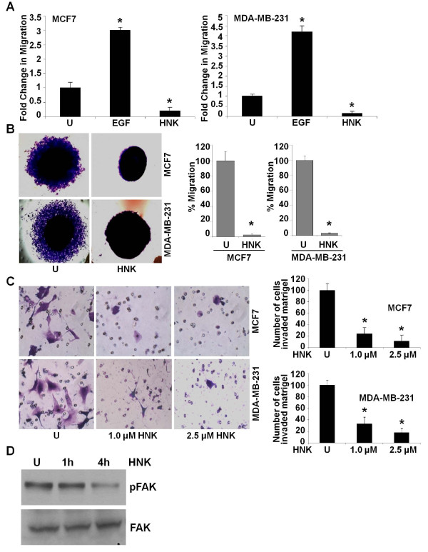
Introduction: Honokiol, a small-molecule polyphenol isolated from magnolia species, is widely known for its therapeutic potential as an antiinflammatory, antithrombosis, and antioxidant agent, and more recently, for its protective function in the pathogenesis of carcinogenesis. In the present study, we sought to examine the effectiveness of honokiol in inhibiting migration and invasion of breast cancer cells and to elucidate the underlying molecular mechanisms.

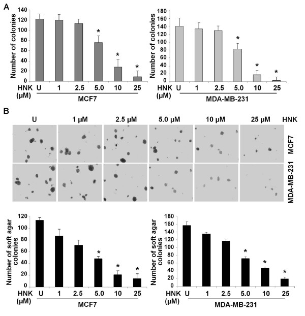
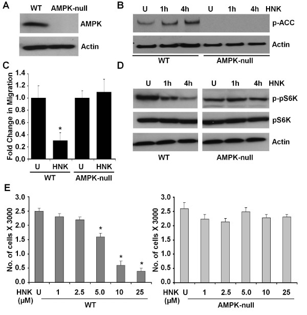
Methods: Clonogenicity and three-dimensional colony-formation assays were used to examine breast cancer cell growth with honokiol treatment. The effect of honokiol on invasion and migration of breast cancer cells was evaluated by using Matrigel invasion, scratch-migration, spheroid-migration, and electric cell-substrate impedance sensing (ECIS)-based migration assays. Western blot and immunofluorescence analysis were used to examine activation of the liver kinase B1 (LKB1)-AMP-activated protein kinase (AMPK) axis. Isogenic LKB1-knockdown breast cancer cell line pairs were developed. Functional importance of AMPK activation and LKB1 overexpression in the biologic effects of honokiol was examined by using AMPK-null and AMPK-wild type (WT) immortalized mouse embryonic fibroblasts (MEFs) and isogenic LKB1-knockdown cell line pairs. Finally, mouse xenografts, immunohistochemical and Western blot analysis of tumors were used.

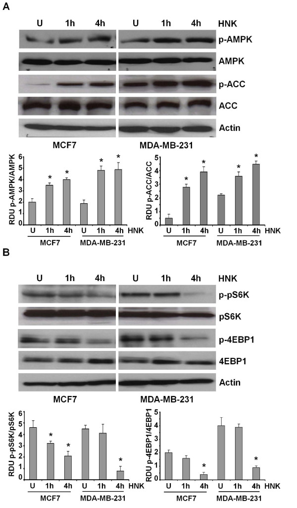
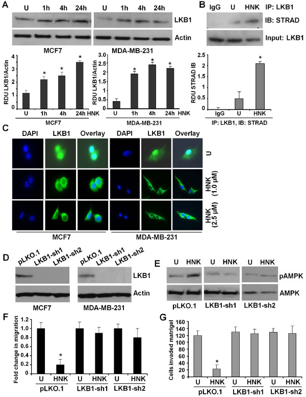
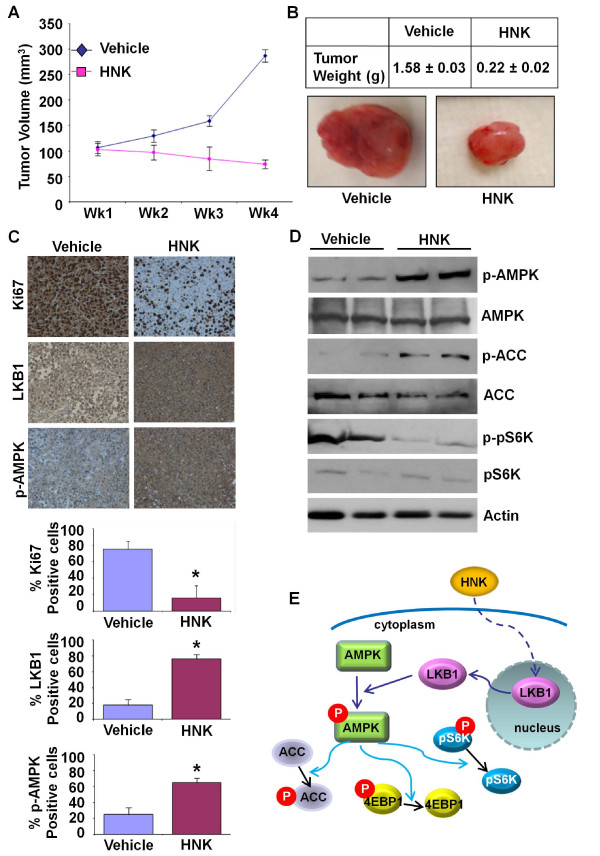
Results: Analysis of the underlying molecular mechanisms revealed that honokiol treatment increases AMP-activated protein kinase (AMPK) phosphorylation and activity, as evidenced by increased phosphorylation of the downstream target of AMPK, acetyl-coenzyme A carboxylase (ACC) and inhibition of phosphorylation of p70S6kinase (pS6K) and eukaryotic translation initiation factor 4E binding protein 1 (4EBP1). By using AMPK-null and AMPK-WT (MEFs), we found that AMPK is required for honokiol-mediated modulation of pACC-pS6K. Intriguingly, we discovered that honokiol treatment increased the expression and cytoplasmic translocation of tumor-suppressor LKB1 in breast cancer cells. LKB1 knockdown inhibited honokiol-mediated activation of AMPK and, more important, inhibition of migration and invasion of breast cancer cells. Furthermore, honokiol treatment resulted in inhibition of breast tumorigenesis in vivo. Analysis of tumors showed significant increases in the levels of cytoplasmic LKB1 and phospho-AMPK in honokiol-treated tumors.

Conclusions: Taken together, these data provide the first in vitro and in vivo evidence of the integral role of the LKB1-AMPK axis in honokiol-mediated inhibition of the invasion and migration of breast cancer cells. In conclusion, honokiol treatment could potentially be a rational therapeutic strategy for breast carcinoma.


Figures

Figure 1
Figure 1
Honokiol inhibits clonogenicity and anchorage-independent growth of breast cancer cells. (a) MCF7 and MDA-MB-231 cells were treated with various concentrations of honokiol (HNK) (as indicated) and subjected to clonogenicity assay. U, untreated cells. Colonies containing > 50 normal-appearing cells were counted. *P < 0.005, compared with untreated controls. (b) Breast cancer cells were subjected to soft-agar colony-formation assay in the presence of various concentrations of honokiol for 3 weeks. Results are expressed as average number of colonies counted (in six microfields). *P < 0.001, compared with untreated controls.
Figure 2
Figure 2
Honokiol inhibits migration and invasion of breast cancer cells. (a) MCF7 and MDA-MB-231 cells were subjected to scratch-migration assay. Culture media were replaced with media containing honokiol (2.5 μM) or untreated media (U). The 100-ng/ml epidermal growth factor (EGF) treatment was used as positive control. The plates were photographed at the identical location of the initial image (0 hours) at 24 hours. The results shown are representative of three independent experiments performed in triplicate. The histogram shows the fold change in migration. *P < 0.01, compared with untreated controls. (b) MCF7 and MDA-MB-231 cells were subjected to spheroid-migration assay. Culture media were replaced with media containing honokiol (2.5 μM) or untreated media (U). The spheroids were photographed 48 hours after treatment. The results shown are representative of three independent experiments performed in triplicate. The histograms show percentage migration. *P < 0.01, compared with untreated controls. (c) MCF7 and MDA-MB-231 cells were cultured in Matrigel invasion chambers followed by treatment with honokiol (HNK, 1.0, 2.5 μM) for 24 hours, as indicated. U, untreated controls. The number of cells that invaded through the Matrigel was counted in five different regions. The slides were blinded to remove counting bias. The histograms show the mean of three independent experiments performed in triplicate. *P < 0.005, compared with untreated controls. (d) Breast cancer cells (MDA-MB-231) were treated with honokiol (HNK, 2.5 μM) for indicated time intervals. U, untreated cells. Total protein was isolated, and equal amounts of proteins were resolved with SDS-PAGE and subjected to immunoblot analysis by using specific antibodies for phosphorylated FAK. The membranes were reblotted by using total FAK antibodies as controls. The blots are representative of multiple independent experiments.
Figure 3
Figure 3
Honokiol activates AMPK and inhibits pS6K and 4EBP1 phosphorylation in breast cancer cells. (a) MCF7 and MDA-MB-231 cells were treated with honokiol (HNK, 2.5 μM) for indicated time intervals. U, untreated cells. Total protein was isolated, and equal amounts of proteins were resolved with SDS-PAGE and subjected to immunoblot analysis by using specific antibodies for phosphorylated AMPK (pAMPK-Thr 172) and phosphorylated ACC (pACC). The membranes were reblotted by using total AMPK and ACC antibodies as controls. The blots are representative of multiple independent experiments. The histogram is the mean of densitometric analysis showing relative density units (RDUs) of the Western blot signal for pAMPK and pACC normalized to total AMPK or ACC in three separate experiments. *P < 0.005, compared with untreated controls. (b) Breast cancer cells were treated with honokiol as in (a) and subjected to immunoblot analysis by using specific antibodies for phosphorylated pS6K (p-pS6K) and phosphorylated 4EBP1 (p-4EBP1). The membranes were reblotted by using total pS6K and p-4EBP1 antibodies as controls. The blots are representative of multiple independent experiments. The histogram is the mean of densitometric analysis showing relative density units (RDUs) of the Western blot signal for p-pS6K and p-4EBP1 normalized to total pS6K or 4EBP1 in three separate experiments. *P < 0.001, compared with untreated controls.
Figure 4
Figure 4
AMPK knockdown abrogates honokiol-mediated increased phosphorylation of ACC, inhibition of phosphorylation of S6K, and inhibition of migration. (a) Immunoblotting for AMPK protein by using lysates from untreated MEFs derived from AMPK-WT (WT) and AMPK-knockout mice (AMPK-null). The blot was stripped and reprobed with anti-actin antibody. (b) WT and AMPK-null MEFs were treated with honokiol (HNK, 2.5 μM) for indicated time intervals. U, untreated cell. Total protein was isolated, and equal amounts of proteins were resolved with SDS-PAGE and subjected to immunoblot analysis by using specific antibodies for phosphorylated ACC (p-ACC). Anti-actin antibody was used as control. (c) WT and AMPK-null MEFs were subjected to scratch-migration assay in the presence (HNK, 2.5 μM) or absence (U) of honokiol. The plates were photographed at the identical location of the initial image (0 hours) at 24 hours. The histogram shows the fold change in migration. *P < 0.001, compared with untreated controls. All the experiments were performed thrice in triplicate. (d) WT and AMPK-null MEFs were treated with honokiol (HNK, 2.5 μM) for indicated time intervals. U, untreated cell. Total protein was isolated, and equal amounts of proteins were resolved with SDS-PAGE and subjected to immunoblot analysis by using specific antibodies for phosphorylated pS6K (p-pS6K). The membranes were reblotted by using total pS6K and actin antibody as control. (e) WT and AMPK-null MEFs were subjected to XTT assay in the presence (HNK) or absence (U) of honokiol, as indicated. The results shown are representative of three independent experiments performed in triplicate. *P < 0.001, compared with untreated controls.
Figure 5
Figure 5
Honokiol increases LKB1 expression, LKB1:STRAD interaction, cytosolic translocation, and depletion of LKB1 abrogates honokiol-mediated modulation of AMPK, inhibition of migration, and invasion of breast cancer cells. (a) MCF7 and MDA-MB-231 cells were treated with 2.5 μM honokiol for indicated time intervals. U, untreated cell. Total protein was isolated, and equal amounts of proteins were resolved with SDS-PAGE and subjected to immunoblot analysis by using specific antibodies for LKB1. The membranes were reblotted by using actin antibody as control. The blots are representative of multiple independent experiments. The histogram is the mean of densitometric analysis showing relative density units (RDUs) of the Western blot signals for LKB1 normalized to actin in three independent experiments. *P < 0.005, compared with untreated controls. (b) MCF7 cells were treated with 2.5 μM honokiol or untreated and subjected to immunoprecipitation assay by using IgG or LKB1 antibodies, as indicated. Immunoprecipitates were analyzed by using anti-STRAD antibodies. The histogram is the mean of densitometric analysis showing relative density units (RDUs) of the Western blot signals for STRAD in three independent experiments. *P < 0.005, compared with untreated controls. (c) MCF7 and MDA-MB-231 cells were treated with honokiol (HNK), and LKB1 protein was analyzed with immunofluorescence by using LKB1 antibody; 4'6-diamidino-2-phenylindole staining was used to determine the nuclear localization. These results are representative of multiple independent experiments. (d) LKB1 was depleted in MCF7 and MDA-MB-231 cells by using two different lentiviral LKB1 short-hairpin RNA (shRNA1 and shRNA2) constructs and a negative control construct that was created in the same vector system (pLKO.1). Stable pools of LKB1-depleted (LKB1shRNA) and vector control (pLKO.1) cells were used for total protein isolation, and equal amounts of proteins were subjected to immunoblot analysis by using specific antibodies for LKB1. Actin was used as control. (e) MDA-MB-231-LKB1shRNA (LKB1-sh1 and LKB1-sh2) and MDA-MB-231-pLKO.1 (pLKO.1) cells were treated with honokiol (HNK, 2.5 μM), and phosphorylation of AMPK was analyzed with Western blot analysis. Total AMPK antibody was used as control. (f) MDA-MB-231-LKB1shRNA (LKB1-sh1 and LKB1-sh2) and MDA-MB-231-pLKO.1 (pLKO.1) cells were grown to confluence, scratched with a pipette tip, and photographed immediately after scratching (0 hours). Culture media were replaced with media containing honokiol (HNK, 2.5 μM) or untreated media (U). The plates were photographed at the identical location of the initial image (0 hours) at 24 hours. The results shown are representative of three independent experiments performed in triplicate. (g) MDA-MB-231-LKB1shRNA (LKB1-sh1 and LKB1-sh2) and MDA-MB-231-pLKO.1 (pLKO.1) cells were cultured in Matrigel invasion chambers followed by treatment with honokiol (HNK, 2.5 μM) for 24 hours. The number of cells that invaded through the Matrigel was counted in five different regions. The slides were blinded to remove counting bias. The result shows the mean of three independent experiments performed in triplicate. *P < 0.005, compared with untreated controls.
Figure 6
Figure 6
Honokiol treatment inhibited breast tumor growth in nude mice. MDA-MB-231 cells-derived tumors were developed in nude mice and treated with vehicle or honokiol (HNK). (a) Tumor growth was monitored by measuring the tumor volume for 4 weeks (eight mice per group). (b) At the end of 6 weeks, tumors were collected, measured, weighed, and photographed. Honokiol treatment inhibited tumor size as compare with vehicle treatment. Average tumor weight and representative tumor images are shown here. (c) Tumor samples were subjected to immunohistochemical analysis by using LKB1, p-AMPK, and Ki67 antibodies. Honokiol (HNK) treatment decreased the expression of Ki-67, increased expression of LKB1 and pAMPK, as compared with vehicle treatment. Bar diagrams show quantitation of protein expression in tumors from vehicle- and honokiol-treated mice. Columns, mean (n = 8); bar, SD. *Significantly different (P < 0.005) compared with control. (d) Tumor lysates (from two different tumors from each set) were subjected to immunoblot analysis by using phospho-AMPK (p-AMPK), AMPK, phospho-ACC (p-ACC), ACC, phospho-pS6K, pS6K antibodies. Actin antibody was used as control. (e) A model of honokiol (HNK)-stimulated AMPK activation in breast cancer cells. Honokiol stimulation induces LKB1 translocation from the nucleus into cytosol and phosphorylates AMPK, leading to increased phosphorylation of ACC and decreased phosphorylation of pS6K and 4EBP1.

Similar articles

Cited by

References

    1. Surh YJ. Cancer chemoprevention with dietary phytochemicals. Nat Rev Cancer. 2003;3:768–780. doi: 10.1038/nrc1189. - DOI - PubMed
    1. Newman DJ, Cragg GM, Snader KM. Natural products as sources of new drugs over the period 1981-2002. J Nat Prod. 2003;66:1022–1037. doi: 10.1021/np030096l. - DOI - PubMed
    1. Fried LE, Arbiser JL. Honokiol, a multifunctional antiangiogenic and antitumor agent. Antioxid Redox Signal. 2009;11:1139–1148. doi: 10.1089/ars.2009.2440. - DOI - PMC - PubMed
    1. Fujita M, Itokawa H, Sashida Y. [Studies on the components of Magnolia obovata Thunb. 3. Occurrence of magnolol and honokiol in M. obovata and other allied plants] Yakugaku Zasshi. 1973;93:429–434. - PubMed
    1. Konoshima T, Kozuka M, Tokuda H, Nishino H, Iwashima A, Haruna M, Ito K, Tanabe M. Studies on inhibitors of skin tumor promotion, IX. Neolignans from Magnolia officinalis. J Nat Prod. 1991;54:816–822. doi: 10.1021/np50075a010. - DOI - PubMed

Publication types

MeSH terms