. 2012 Mar 13;106(6):1205-13.
doi: 10.1038/bjc.2012.26. Epub 2012 Feb 21.

Creation of immortalised epithelial cells from ovarian endometrioma

Affiliations

Creation of immortalised epithelial cells from ovarian endometrioma

Y Bono et al. Br J Cancer. .

Abstract

Background: Epithelial cells of endometriotic tissues are difficult to propagate in vitro as experimental material is scarce owing to their limited life span. However, there is an increasing concern regarding their malignant transformation in ovaries. The present study sought to generate their stable culture system.

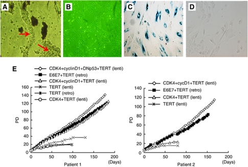
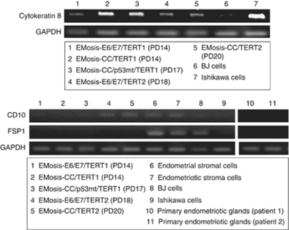
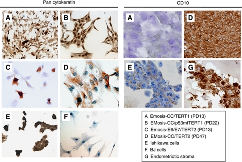
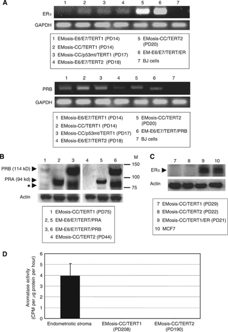
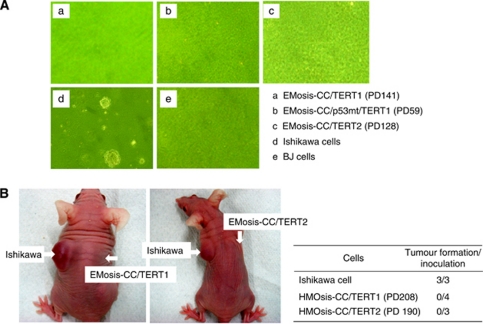
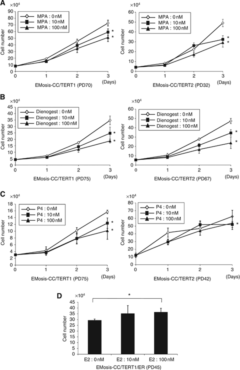
Methods and results: Purified epithelial cells isolated from ovarian endometriomas using microscopic manipulation were successfully immortalised by combinatorial transfection of human cyclinD1, cdk4 and human telomerase reverse transcriptase (hTERT) genes, whereas the introduction of hTERT alone, or together with cdk4, was insufficient for immortalisation, leading to cellular senescence. We confirmed stable cytokeratin expression in the immortalised cells, proving their epithelial origin. These cells expressed progesterone receptor B and showed significant growth inhibition by various progestins. Oestrogen receptor (ER) expression was detected in these cells, albeit at low levels. Additional overexpression of ERα generated stable cells with oestrogen-dependent growth activation. Soft-agar colony formation assay and nude mice xenograft experiments demonstrated that these cells, even those with additional inactivation of p53, did not have transformed phenotypes.

Conclusion: We for the first time generated immortalised epithelial cells from ovarian endometrioma that retained sex steroid responsiveness. These cells are invaluable tools not only for the consistent in vitro work but also for the study of molecular pathogenesis or carcinogenesis of endometriosis.


Conflict of interest statement

The authors declare no conflict of interest.

Figures

Figure 1
Figure 1
Morphological characteristics and proliferative life span of epithelial cells from ovarian endometrioma transfected with various genetic factors. (A) Phase contrast image of glandular clusters isolated from ovarian endometrioma tissues. Individual clusters were directly picked up, one by one, using microscopic manipulation and were transfected with various genetic factors. Glandular clusters are shown in arrows. (B) Phase contrast image of representative isolated clones (EMosis-CC /TERT-1 cells) cultured on plastic dishes are shown. (C) β-gal staining of cells from patient 1 transfected with hTERT alone (population doubling (PD): 20). (D) β-gal staining of EMosis-CC /TERT-1 cells (PD: 100). (E) The growth characteristics of transfected cells are represented as a growth curve. The genetic factors introduced are shown. Abbreviations: lenti=lentiviral vectors; retro=retroviral vectors.
Figure 2
Figure 2
RT–PCR analysis of the expression of epithelial and stromal markers in immortalised epithelial cells from ovarian endometrioma. The expression of cytokeratin 8, CD10 and FSP1 in endometriotic epithelial cells immortalised by various genetic factors was examined using RT–PCR. Ishikawa and BJ cells were used as controls for epithelial and fibroblast cells, respectively. Primary endometriotic glandular cells without transfection, isolated from the ovarian endometrioma of patient 1 or 2, were used as negative controls for CD10 or FSP1 expression. GAPDH expression was assayed as a loading control.
Figure 3
Figure 3
Immunocytochemical analyses of cytokeratin and CD10 expression in immortalised epithelial cells from ovarian endometrioma. The expression of cytokeratin and CD10 in endometriotic epithelial cells that were immortalised by various genetic factors and cultured on LAB TEK chamber slides was examined using immunocytochemistry. Ishikawa and BJ cells were used as controls for epithelial and fibroblast cells, respectively. Primary stromal cells without transfection, isolated from the ovarian endometrioma of another patient, were used as a positive control for CD10.
Figure 4
Figure 4
Sex steroid-receptor expression in, and aromatase activity of, immortalised epithelial cells from ovarian endometrioma. (A) RT–PCR analysis of expression of the oestrogen receptor α (ERα) or the progesterone receptor B (PRB). EM-E6/E7/TERT/ER cells are immortalised endometrial epithelial cells in which ERα cDNAs were stably transfected and were used as a positive control for ERα. EM-E6/E7/TERT/PRB cells are immortalised endometrial epithelial cells in which PRB cDNAs were stably transfected. Because our primer sets for PRB were designed to amplify the sequences containing PRB gene promoter in order to distinguish from PRA transcript, they can detect only intrinsic PRB mRNA but not extrinsic, overexpressed PRB mRNA that lacks promoter sequences. The weak PRB band in EM-E6/E7/TERT/PRB cells is therefore derived from intrinsic PRB. BJ cells were used as a negative control for ERα and PRB expression. GAPDH was used as a loading control. (B) Western blot analysis of expression of the progesterone receptor. EM-E6/E7/TERT/PRA or EM-E6/E7/TERT/PRB cells are immortalised endometrial epithelial cells in which PRA or PRB cDNAs were stably transfected and were used as a positive control for PRA or PRB expressions, respectively. Although EM-E6/E7/TERT/PRA cells showed a clear PRA band by western blotting (94 kDa), EM-E6/E7/TERT/PRB cells displayed two bands; one band was of the expected size of intact PRB (114 kDa); the other band was located just below the PRA band (identified by the symbol: ★) and was not a PRA band but a degraded PRB band, which was confirmed by another western blot analysis using a PRB-specific antibody (data not shown). EMosis-CC/TERT1 cells exhibited a weak, but distinct, PRB band but not a PRA band. M: protein weight marker. (C) Western blot analysis of expression of the ER. There was no detectable protein expression of ERα in EMosis-CC/TERT1 or EMosis-CC/TERT2 cells. EMosis-CC/TERT1/ER cells, generated by the introduction of ERα cDNA into EMosis-CC/TERT1 cells, were confirmed to have significant ERα expression. MCF7 cells were used as a positive control of ERα expression. (D) Analysis of aromatase activity using a tritiated water assay. Primary endometriotic stromal cells isolated from the ovarian endometrioma of another patient were used as a positive control of aromatase activity. Both EMosis-CC/TERT1 and EMosis-CC/TERT2 cells lacked aromatase activity.
Figure 5
Figure 5
Analysis of the transformed phenotypes of immortalised epithelial cells from ovarian endometrioma. (A) Anchorage-independent growth was examined using a soft-agar colony formation assay. A total of 2 × 10⁵ Ishikawa cells or immortalised cells were seeded onto soft agar and colonies >0.2 mm were counted after incubation for 2 weeks. Ishikawa and BJ cells were used as a positive and negative control for colony formation, respectively. (B) In vivo growth was examined using a tumour formation assay in nude mouse. Immortalised epithelial cells from ovarian endometrioma were resuspended in growth media (108 cells per ml) and were subcutaneously injected (0.1 ml) into the base of the bilateral flank of female BALB/c nu/nu mice (age range 7–9 weeks, SLC). Tumour growth was monitored weekly until confirmed tumours were visualised or at least for 2 months unless tumour formation was detected.
Figure 6
Figure 6
Effect of progestin and oestrogen on the growth of immortalised epithelial cells from ovarian endometrioma. EMosis-CC/TERT1 or EMosis-CC/TERT2 cells were seeded on six-well dishes and were treated with or without MPA (A), dienogest (B) or progesterone (P4) (C) at a concentration of 10 or 100 nM for the indicated number of days. Cell growth was monitored by counting cell numbers. Data are presented as means±s.d. of three independent experiments. *P<0.05. (D) EMosis-CC/TERT1/ER cells were generated by the introduction of ERα cDNA into EMosis-CC/TERT1 cells and confirmed to have significant ERα expression (Figure 4C). EMosis-CC/TERT1/ER cells were seeded on six-well dishes and were treated with or without MPA estradiol (E2) at a concentration of 10 or 100 nM for the indicated number of days. Cell growth was monitored by counting cell numbers on day 5 after treatment. Data are presented as means±s.d. of three independent experiments. *P<0.05.

Similar articles

Cited by

References

    1. Anglesio MS, Carey MS, Köbel M, Mackay H, Huntsman DG, Vancouver Ovarian Clear Cell Symposium Speakers (2010) Clear cell carcinoma of the ovary: A report from the first Ovarian Clear Cell Symposiumh. Gynecol Oncol 121: 407–415 - PubMed
    1. Attia GR, Zeitoun K, Edwards D, Johns A, Carr BR, Bulun SE (2000) Progesterone receptor isoform A but not B is expressed in endometriosis. J Clin Endocrinol Metab 85: 2897–2902 - PubMed
    1. Bulun SE, Mahendroo MS, Simpson ER (1993) Polymerase chain reaction amplification fails to detect aromatase cytochrome P450 transcripts in normal human endometrium or decidua. J Clin Endocrinol Metab 76: 1458–1463 - PubMed
    1. Bulun SE, Yang S, Fang Z, Gurates B, Tamura M, Zhou J Sebastian S (2001) Role of aromatase in endometrial disease. J Steroid Biochem Mol Biol 79: 19–25 - PubMed
    1. Bulun SE, Cheng YH, Yin P, Imir G, Utsunomiya H, Attar E, Innes J, Julie, Kim J (2006) Progesterone resistance in endometriosis: link to failure to metabolize estradiol. Mol Cell Endocrinol 248: 94–103 - PubMed

Publication types

MeSH terms