. 2012 Mar;122(3):821-32.
doi: 10.1172/JCI61014. Epub 2012 Feb 22.

Partial MCM4 deficiency in patients with growth retardation, adrenal insufficiency, and natural killer cell deficiency

Affiliations

Partial MCM4 deficiency in patients with growth retardation, adrenal insufficiency, and natural killer cell deficiency

Laure Gineau et al. J Clin Invest. 2012 Mar.

Abstract

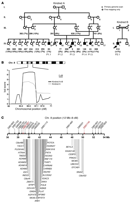
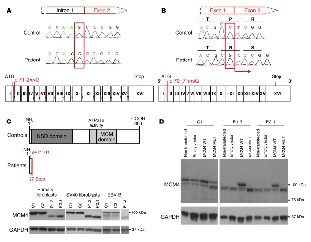
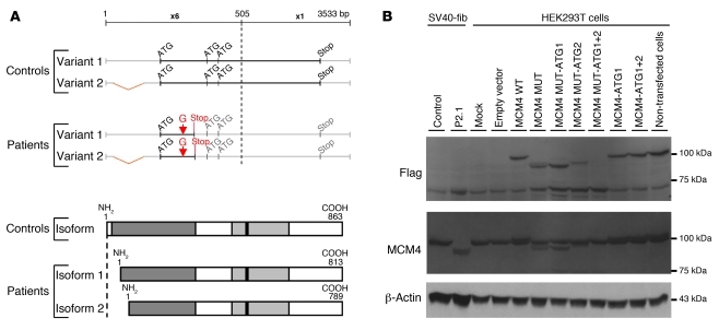
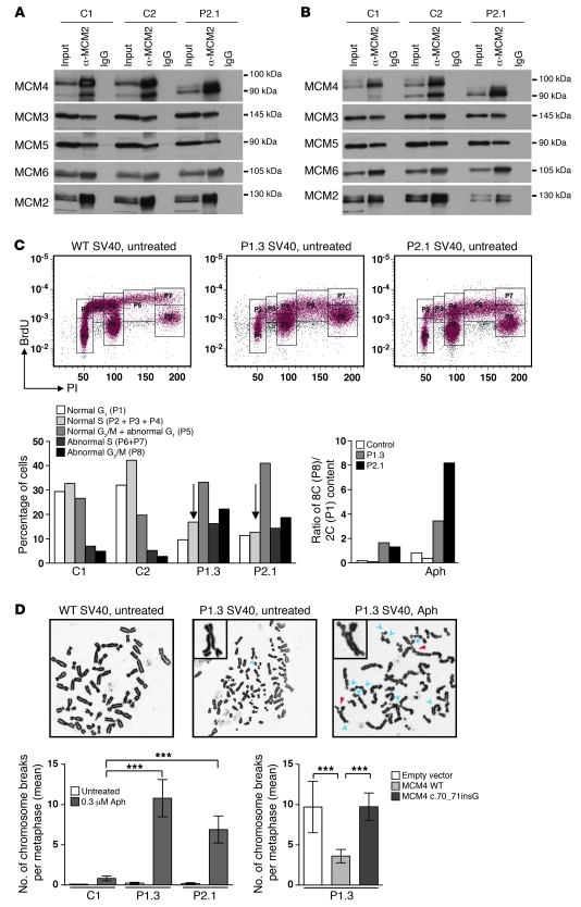
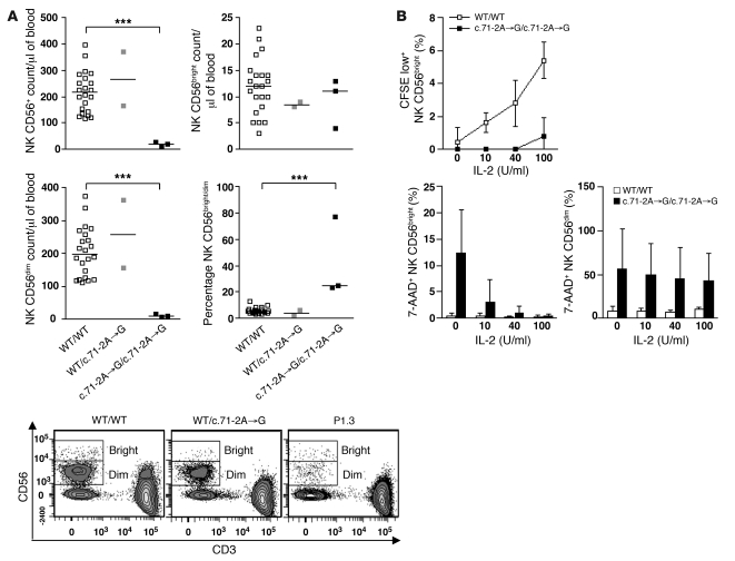
Natural killer (NK) cells are circulating cytotoxic lymphocytes that exert potent and nonredundant antiviral activity and antitumoral activity in the mouse; however, their function in host defense in humans remains unclear. Here, we investigated 6 related patients with autosomal recessive growth retardation, adrenal insufficiency, and a selective NK cell deficiency characterized by a lack of the CD56(dim) NK subset. Using linkage analysis and fine mapping, we identified the disease-causing gene, MCM4, which encodes a component of the MCM2-7 helicase complex required for DNA replication. A splice-site mutation in the patients produced a frameshift, but the mutation was hypomorphic due to the creation of two new translation initiation methionine codons downstream of the premature termination codon. The patients' fibroblasts exhibited genomic instability, which was rescued by expression of WT MCM4. These data indicate that the patients' growth retardation and adrenal insufficiency likely reflect the ubiquitous but heterogeneous impact of the MCM4 mutation in various tissues. In addition, the specific loss of the NK CD56(dim) subset in patients was associated with a lower rate of NK CD56(bright) cell proliferation, and the maturation of NK CD56(bright) cells toward an NK CD56(dim) phenotype was tightly dependent on MCM4-dependent cell division. Thus, partial MCM4 deficiency results in a genetic syndrome of growth retardation with adrenal insufficiency and selective NK deficiency.


Figures

Figure 1
Figure 1. Linkage analysis of NK cell deficiency in humans.
(A) Pedigrees of the two families. Generations are designated by Roman numerals I–IV. Patients with low counts of NK cells (P1.1, P1.2, P1.3, P1.4, P1.5, and P2.1) are represented by black symbols. The index case is indicated by an arrow. All other family members with normal NK cell counts are indicated by white symbols. A star indicates that the individual has been genotyped for all the microsatellites considered, whereas a circle indicates that genotyping has been carried out only for the 8p12–q12.2 region. Absolute numbers (per mm3 of whole blood) and percentages (% of lymphocytes) of NK cells are indicated for each individual, for the first whole-blood sample analyzed. (B) Multipoint linkage analysis of chromosome region 8p12–q12.2 by homozygosity mapping. Microsatellite positions are indicated in cM. In total, 9 microsatellites were genotyped between D8S1821 and D8S1745. The gray line represents kindred A lod score, and the black line represents the combined lod score for kindreds A and B. (C) Schematic representation of the candidate region (8p11.23–q11.21). The region of interest was delineated between microsatellites D8S1821 and D8S1745. This region is 12 Mb long and contains 1 microRNA and 45 predicted protein-coding genes.
Figure 2
Figure 2. The MCM4 mutation is not associated with a loss of expression.
(A) Automated sequencing profiles showing the homozygous MCM4 c.70_71insG mutation in genomic DNA extracted from EBV-B cells of a patient and a WT control. Bottom: Schematic diagram of the structure of the MCM4 gene, consisting of 16 or 17 exons (Roman numerals), indicating the position of the mutation, which affects the acceptor splice site of intron 1. (B) Sequencing profile of a patient and a control indicating the insertion of a G nucleotide in the cDNA extracted from EBV-B cells. The position of the mutation is shown on a diagram of the MCM4 gene. The dotted lines represent the two mRNA transcripts produced from MCM4. The homozygous mutation leads to the insertion of an additional nucleotide between exons 1 and 2. (C) A schematic diagram of the MCM4 protein, which has an N-terminal serine/threonine-rich domain (dark gray) and a conserved MCM domain (light gray) including an ATP-binding site (black) toward its C terminus. The homozygous mutation results in a frameshift, creating a premature stop codon in exon 2. Bottom: Western blot analysis of MCM4 on total protein extracts from primary fibroblasts and SV40 fibroblasts from P1.3 and P2.1 and EBV-B cells from P1.2, controls. A polyclonal MCM4 antibody was used. (D) Complementation, by lentiviral particles, of SV40 fibroblasts from the controls and patients, with an empty pTRIP vector, an MCM4 WT vector, and the MCM4 c.71-2A→G mutation (MCM4 MUT). MCM4 was detected with a polyclonal antibody. The empty vector and non-transfected cells were used as a negative transfection control. In C and D, GAPDH was used as a loading control.
Figure 3
Figure 3. Characterization of the MCM4 isoforms detected in the cells of the patient tested.
Reinitiation of MCM4 protein translation. (A) Schematic diagram of the two potential reinitiation sites after the premature STOP codon and the corresponding isoforms of MCM4. Two ATG codons, at positions 51 and 75, are in the same open reading frame as the premature stop codon, leading to the production of two new isoforms, of 813 and 789 amino acids, respectively. The 5′ part of the MCM4 sequence is enlarged (×6) and shown at greater magnification than the 3′ part of the MCM4 sequence (×1). The two variants of MCM4 mRNA from the NCBI database are shown (NM 005914.3 and NM 182746.2). (B) MCM4 protein levels, in HEK293T cells, following transient transfection with a C-terminal Flag-tagged pCMV6 empty vector or pCMV6 MCM4 WT, pCMV6 MCM4 MUT, pCMV6 MCM4 MUT-ATG1, pCMV6 MCM4 MUT-ATG2, pCMV6 MCM4 MUT-ATG1+2, pCMV6 MCM4-ATG1, and pCMV6 MCM4-ATG1+2 vectors, were assessed by Western blotting of total protein extracts from each transfection with antibodies against Flag and against the MCM4 protein. Total protein extracts from non-transfected control SV40 fibroblast (SV40-Fib) cell lines from a control and patient P2.1 were used as a positive control. An antibody against β-actin was used as a loading control.
Figure 4
Figure 4. Functionality of the MCM4 isoforms detected in the cells of the patient.
(A) The Triton X–extractable fraction from patient and control SV40 fibroblasts was subjected to immunoprecipitation with a monoclonal antibody against MCM2. Protein extracts and immunoprecipitates were analyzed by immunoblotting with antibodies against MCM4, MCM3, MCM5, and MCM6. MCM2 was used as a loading control. (B) The chromatin-bound fraction was subjected to immunoprecipitation with a monoclonal antibody against MCM2. This procedure was carried out on the DNase I–extracted fractions of both control cells and cells from the patient. Protein extracts and immunoprecipitates were analyzed by immunoblotting with antibodies against MCM4, MCM3, MCM5, and MCM6. MCM2 was used as a loading control. Immunoprecipitation with IgG was used as a negative control. (C) Representative flow cytometry plots of the cell cycle of SV40 fibroblasts from controls and patients. Control (left), P1.3 (middle), and P2.1 (right) cell cycles in the absence of treatment. Transformation of cells with SV40 T antigen causes increased ploidy of all cells (59). P1 corresponds to normal G1 phase, P2+P3+P4 correspond to normal S phase, P5 corresponds to normal G2 phase plus abnormal G1 or failed mitosis, P6+P7 correspond to re-replication S phase, and P8 corresponds to 8C (P8) DNA content. Patients’ SV40 fibroblasts with or without 0.3 μM aphidicolin (Aph) treatment. (D) Representative chromosome spreads of chromosome breaks induced with or without aphidicolin. A WT metaphase chromosome without aberrations (left); a P1.3 metaphase with some aberrations indicated by blue arrowhead (middle); and a P1.3 metaphase after aphidicolin treatment, with chromosomal aberrations indicated by arrowheads (right). Blue arrowheads indicate chromatid breaks, and red arrowheads indicate chromosome exchanges. Bottom: Chromosome breaks (mean) per metaphase in P1.3 and P2.1 SV40 fibroblasts and in control SV40 fibroblasts. Complementation by lentiviral transduction with the WT MCM4 allele, the empty vector, or the c.70_71insG allele in P1.3 SV40 fibroblasts. Error bars indicate SEM. ***P < 0.0005, Student’s t test.
Figure 5
Figure 5. Homozygous MCM4 mutation and specific NK CD56dim deficiency.
(A) Quantitation, by flow cytometry, of peripheral total NK cells and of the CD56bright and CD56dim NK cell subsets in controls (n = 22), heterozygous subjects (n = 2), and homozygous patients (P1.1, P1.2, and P1.3). Horizontal bars represent medians. ***P < 0.0005, Student’s t test. Bottom: Representative flow cytometry plots of a homozygous WT sibling, a heterozygous sibling, and one patient. (B) PBMCs from 6 independent healthy controls and from one patient (P1.3) tested in 2 independent experiments were stained with CFSE and stimulated for 72 hours with various doses of IL-2. Apoptosis was assessed on NK subsets, by 7-AAD staining. Error bars indicate SD.

Comment in

References

    1. Vivier E, et al. Innate or adaptive immunity? The example of natural killer cells. Science. 2011;331(6013):44–49. - PMC - PubMed
    1. Moretta L, Moretta A. Unravelling natural killer cell function: triggering and inhibitory human NK receptors. Embo J. 2004;23(2):255–259. doi: 10.1038/sj.emboj.7600019. - DOI - PMC - PubMed
    1. French AR, Yokoyama WM. Natural killer cells and viral infections. Curr Opin Immunol. 2003;15(1):45–51. - PubMed
    1. Orr MT, Lanier LL. Natural killer cell education and tolerance. Cell. 2010;142(6):847–856. doi: 10.1016/j.cell.2010.08.031. - DOI - PMC - PubMed
    1. Cerwenka A, Lanier LL. Natural killer cells, viruses and cancer. Nat Rev Immunol. 2001;1(1):41–49. doi: 10.1038/35095564. - DOI - PubMed

Publication types

MeSH terms