

Thymic stromal lymphopoietin (TSLP)-mediated dermal inflammation aggravates experimental asthma
- PMID: 22354320
- PMCID: PMC3328620
- DOI: 10.1038/mi.2012.14
Thymic stromal lymphopoietin (TSLP)-mediated dermal inflammation aggravates experimental asthma
Erratum in
- Mucosal Immunol. 2012 Jul;5(4):468
Abstract
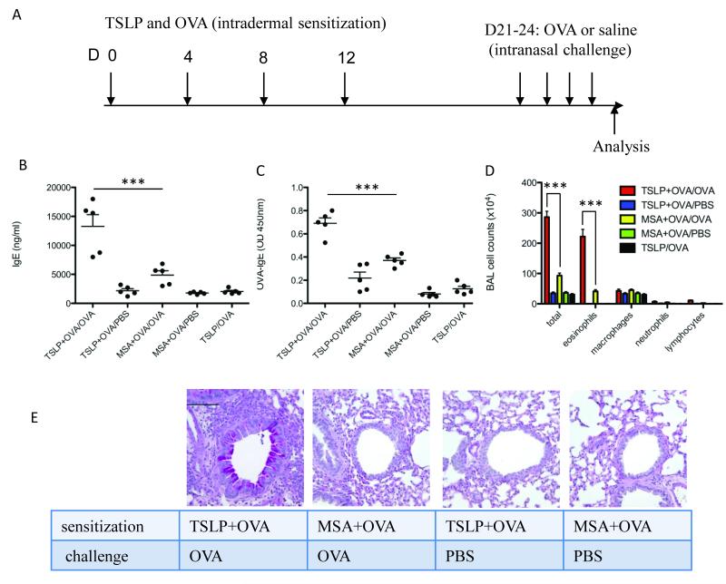
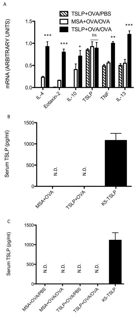
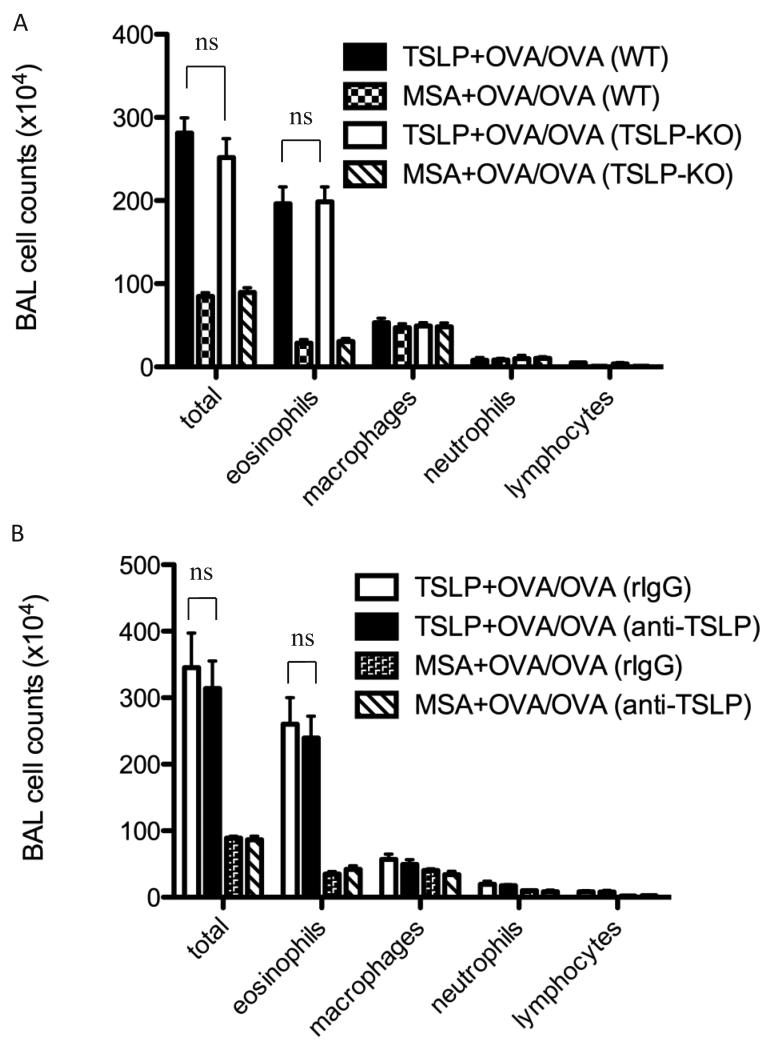
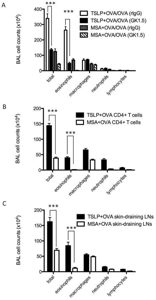
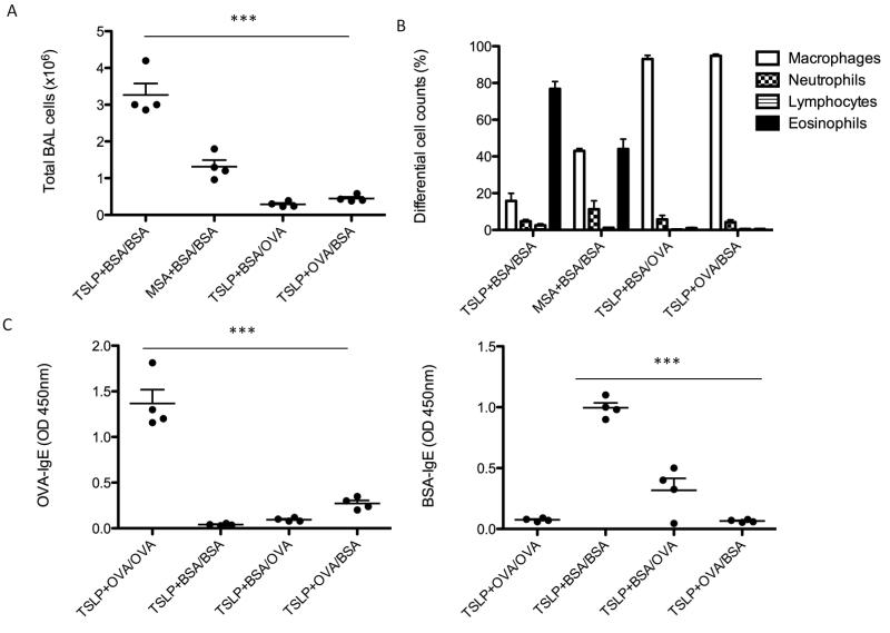
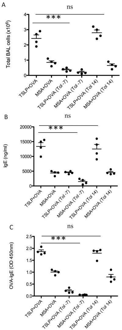
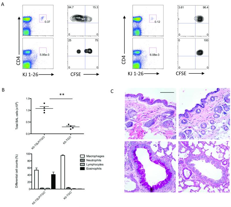
Individuals with one atopic disease are far more likely to develop a second. Approximately half of all atopic dermatitis (AD) patients subsequently develop asthma, particularly those with severe AD. This association, suggesting a role for AD as an entry point for subsequent allergic disease, is a phenomenon known as the "atopic march." Although the underlying cause of the atopic march remains unknown, recent evidence suggests a role for the cytokine thymic stromal lymphopoietin (TSLP). We have established a mouse model to determine whether TSLP plays a role in this phenomenon, and in this study show that mice exposed to the antigen ovalbumin (OVA) in the skin in the presence of TSLP develop severe airway inflammation when later challenged with the same antigen in the lung. Interestingly, neither TSLP production in the lung nor circulating TSLP is required to aggravate the asthma that was induced upon subsequent antigen challenge. However, CD4 T cells are required in the challenge phase of the response, as was challenge with the sensitizing antigen, demonstrating that the response was antigen specific. This study, which provides a clean mouse model to study human atopic march, indicates that skin-derived TSLP may represent an important factor that triggers progression from AD to asthma.
Figures
Similar articles
-
Thymic stromal lymphopoietin-activated basophil promotes lung inflammation in mouse atopic march model.Front Immunol. 2025 May 15;16:1573130. doi: 10.3389/fimmu.2025.1573130. eCollection 2025. Front Immunol. 2025. PMID: 40443683 Free PMC article.
-
TSLP produced by keratinocytes promotes allergen sensitization through skin and thereby triggers atopic march in mice.J Invest Dermatol. 2013 Jan;133(1):154-63. doi: 10.1038/jid.2012.239. Epub 2012 Jul 26. J Invest Dermatol. 2013. PMID: 22832486
-
Skin thymic stromal lymphopoietin promotes airway sensitization to inhalant house dust mites leading to allergic asthma in mice.Allergy. 2012 Aug;67(8):1078-82. doi: 10.1111/j.1398-9995.2012.02857.x. Epub 2012 Jun 12. Allergy. 2012. PMID: 22687045
-
Thymic stromal lymphopoietin: a promising therapeutic target for allergic diseases.Int Arch Allergy Immunol. 2013;160(1):18-26. doi: 10.1159/000341665. Epub 2012 Aug 30. Int Arch Allergy Immunol. 2013. PMID: 22948028 Review.
-
Thymic stromal lymphopoietin:a potential therapeutic target for allergy and asthma.Curr Allergy Asthma Rep. 2006 Sep;6(5):372-6. doi: 10.1007/s11882-996-0006-7. Curr Allergy Asthma Rep. 2006. PMID: 16899198 Review.
Cited by
-
Niche-Specific Factors Dynamically Regulate Sebaceous Gland Stem Cells in the Skin.Dev Cell. 2019 Nov 4;51(3):326-340.e4. doi: 10.1016/j.devcel.2019.08.015. Epub 2019 Sep 26. Dev Cell. 2019. PMID: 31564613 Free PMC article.
-
New Cytokines in the Pathogenesis of Atopic Dermatitis-New Therapeutic Targets.Int J Mol Sci. 2018 Oct 9;19(10):3086. doi: 10.3390/ijms19103086. Int J Mol Sci. 2018. PMID: 30304837 Free PMC article. Review.
-
Thymic stromal lymphopoietin, skin barrier dysfunction, and the atopic march.Ann Allergy Asthma Immunol. 2021 Sep;127(3):306-311. doi: 10.1016/j.anai.2021.06.004. Epub 2021 Jun 19. Ann Allergy Asthma Immunol. 2021. PMID: 34153443 Free PMC article. Review.
-
Effects of microRNA-19b on airway remodeling, airway inflammation and degree of oxidative stress by targeting TSLP through the Stat3 signaling pathway in a mouse model of asthma.Oncotarget. 2017 Jul 18;8(29):47533-47546. doi: 10.18632/oncotarget.17258. Oncotarget. 2017. PMID: 28472780 Free PMC article.
-
Veronica persica Ethanol Extract Ameliorates Dinitrochlorobenzene-Induced Atopic Dermatitis-like Skin Inflammation in Mice, Likely by Inducing Nrf2/HO-1 Signaling.Antioxidants (Basel). 2023 Jun 13;12(6):1267. doi: 10.3390/antiox12061267. Antioxidants (Basel). 2023. PMID: 37371997 Free PMC article.
References
-
- Bieber T. Atopic dermatitis. N. Engl. J. Med. 2008;358:1483–1494. - PubMed
-
- Spergel JM, Paller AS. Atopic dermatitis and the atopic march. J. Allergy Clin. Immunol. 2003;112:S118–S127. - PubMed
-
- Beck LA, Leung DY. Allergen sensitization through the skin induces systemic allergic responses. J. Allergy Clin. Immunol. 2000;106:S258–S263. - PubMed
-
- Friend SL, et al. A thymic stromal cell line supports in vitro development of surface IgM+ B cells and produces a novel growth factor affecting B and T lineage cells. Exp. Hematol. 1994;22:321–328. - PubMed
Publication types
MeSH terms
Substances
Grants and funding
LinkOut - more resources
Full Text Sources
Medical
Molecular Biology Databases
Research Materials
