. 2012 Apr 15;72(8):2111-9.
doi: 10.1158/0008-5472.CAN-11-2209. Epub 2012 Feb 21.

Deficiency in mammalian histone H2B ubiquitin ligase Bre1 (Rnf20/Rnf40) leads to replication stress and chromosomal instability

Affiliations

Deficiency in mammalian histone H2B ubiquitin ligase Bre1 (Rnf20/Rnf40) leads to replication stress and chromosomal instability

Sophia B Chernikova et al. Cancer Res. .

Abstract

Mammalian Bre1 complexes (BRE1A/B (RNF20/40) in humans and Bre1a/b (Rnf20/40) in mice) function similarly to their yeast homolog Bre1 as ubiquitin ligases in monoubiquitination of histone H2B. This ubiquitination facilitates methylation of histone H3 at K4 and K79, and accounts for the roles of Bre1 and its homologs in transcriptional regulation. Recent studies by others suggested that Bre1 acts as a tumor suppressor, augmenting expression of select tumor suppressor genes and suppressing select oncogenes. In this study, we present an additional mechanism of tumor suppression by Bre1 through maintenance of genomic stability. We track the evolution of genomic instability in Bre1-deficient cells from replication-associated double-strand breaks (DSB) to specific genomic rearrangements that explain a rapid increase in DNA content and trigger breakage-fusion-bridge cycles. We show that aberrant RNA-DNA structures (R-loops) constitute a significant source of DSBs in Bre1-deficient cells. Combined with a previously reported defect in homologous recombination, generation of R-loops is a likely initiator of replication stress and genomic instability in Bre1-deficient cells. We propose that genomic instability triggered by Bre1 deficiency may be an important early step that precedes acquisition of an invasive phenotype, as we find decreased levels of BRE1A/B and dimethylated H3K79 in testicular seminoma and in the premalignant lesion in situ carcinoma.


Figures
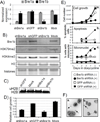
Fig. 1
Fig. 1. Depletion of Bre1a or Bre1b impairs cell growth
(A) qRT-PCR showing reduced mRNA levels of Bre1a or Bre1b in the knockdowns of Bre1a or Bre1b, respectively. (B) Knockdown of Bre1a or Bre1b results in decrease in H3K4me3 and H3K79me2. (C) Depletion of Bre1a or Bre1b decreases monoubiquitination of histone H2B. (D) Depletion of Bre1a and Bre1b results in slow growth of RIF-1 cells. (E) Conditional expression of Bre1b shRNA in mouse RIF-1 cells results in (a) slow growth, (b) increased apoptosis (as measured by annexin-FITC staining), and (c) increased induction of micronuclei. Error bars are standard errors of the mean. Experiments were repeated more than three times. (+) and (−) show presence or absence of doxycycline in the medium. (F) Representative microphotograph showing micronuclei in RIF-1 cells after knockdown of Bre1a (panel a) or Bre1b (panel b).
Fig. 2
Fig. 2. Evidence for ongoing genomic instability in Bre1-depleted cells
(A, B) Bre1b depletion leads to increased frequency of micronucleus (MN) formation. Note different levels of Bre1b, uH2B and H3K79me2 in two doxycycline-inducible cell lines, shBre1b1 and shBre12. shBre1b1, a Bre1b shRNA doxycycline-inducible cell line shows a partial Bre1b knockdown even in the absence of doxycycline. shBre1b2 is derived from early passage shBre1b1 cells line by increasing the stringency of blasticidin selection (from 5 ug/ml to 10 ug/ml). The (+) and (−) show presence or absence of doxycycline in the medium. (C) Anaphase bridges in Bre1b-depleted cells contain telomeric sequences (arrows). (D) Bre1b-depleted cells are characterized by appearance of aberrations with amplified centromeric heterochromatin (stained by DAPI). Shown is a Robertsonian-like Translocation with Amplified Centromeric Heterochromatin (RTCH). (E) Long-term partial knockdown of Bre1b in the shBre1b1 cell line leads to multiple chromosomal abnormalities, which increase in frequency and complexity under. Abbreviations: CA – chromatid-type aberrations; RT – classic Robertsonian Translocations; RTCH – Robertsonian-like Translocations involving Centromeric Heterochromatin; ACH - complex aberrations involving multiple amplification of heterochromatin. Note that RTCH frequencies are much higher in shBre1b1 cells at later passages. Significance analysis is provided in Table S5. (F) Representative examples of observed chromosome aberrations: panels a and b – chromatid-type (CA) aberrations, panels c, d and e – complex aberrations involving heterochromatin (ACH). Arrowhead points to RTCH in (F) panel e.
Fig. 3
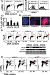
Fig. 3. Bre1 knockdown leads to increase in double-strand breaks and to replication stress
(A) FACS analysis shows that loss of Bre1b elevates γH2AX. A representative experiment is shown. Increase in γH2AX signal results from an increase in intensity of γH2AX foci (B) and from increased number of cells counted as γH2AX-positive (C). (D). Panel (a) shows cells with low γH2AX signal and panels (b) and (c) show cells with more intense γH2AX foci after Bre1 knockdown. Panel (c) shows a pre-apoptotic cell with very bright γH2AX signal. (E) Bre1-deficient cells show more cells with high γH2AX signal specifically in S-phase. For quantitation purposes FACS settings were adjusted so that the cells were in the same location on the plots. Note that in the corresponding Western blot % of γH2AX –positive cells is inversely proportional the levels of uH2B in the Bre1a or Bre1b knockdowns. (F) 24 hour treatment with hydroxyurea leads to different distribution of γH2AX signal in the Bre1 knockdown cells. (+) and (−) show presence or absence of doxycycline in medium. Experiments were repeated more than three times.
Fig. 4
Fig. 4. Defect in mRNA processing contributes to double-strand break formation in Bre1-depleted cells
(A) Classification enrichment in gene sets down-regulated and upregulated after Bre1 knockdown. Pathway/Process enrichment analysis was performed using DAVID bioinformatics database (Supplemental Methods). The statistical threshold was applied for – log(p=0.05) shown in dashed line. (B) Overexpression of RNase H1 in the shBre1b cells results in reduction of cells with γH2AX foci. (C) Overexpression of RNase H1 in Bre1b-depleted cells reduces number of γH2AX foci per cell. (D) A representative photograph of an experiment from (C). The cells were transfected with GFP-RNase H1 or GFP-Nuc (control) on day 3 after the induction of knockdown and were analysed on day 6 after the induction of knockdown. In (B) the GFP-expressing cells were FACS sorted on day 5 after the induction of knockdown and plated for analysis to be performed next day. The FACS sorting experiments were performed twice. In (C) cells transfected with GFP-RNase H1 were analysed without prior sorting. Asterisks denote a significant difference from cells with Bre1 depletion, but not expressing RNase H1 (*** correspond to p<0.001, t-test).
Fig. 5
Fig. 5. Loss of BRE1A/B is associated with development of seminoma
(A) Bre1b is highly expressed in mouse testis. (B) Bre1b levels are the highest in meiotic prophase cells (depicted by arrows). Staining of Bre1b, H3K79me2, H3K4me3 and of total histone H3 is shown. (C) Protein levels of BRE1B, H3K79me and H3K4me3 are lower in seminoma compared to normal testis. (D) H3K79me2 is significantly lower in seminoma compared to normal testis and non-seminomatous cancers of testis. (E) Representative pictures show that loss of Bre1A/B and H3K79me2 occur early in seminoma development. Normal testis, CIS – carcinoma in situ, and seminoma sections are taken from same patients. Arrows point to CIS. Asterisks denote a significant difference from normal tissue section (*** correspond to p<0.001, and ** correspond to p<0.01, t-test).
Fig. 6
Fig. 6. A model depicting sources of genomic instability in Bre1-deficient cells
Loss of Bre1 leads to increase in DSBs due to defects in homologous recombination and in the processing of canonical histone mRNAs. Incorrect processing of replication-dependent histone mRNA 3’-ends facilitates co-transcriptional formation of R-loops, which block replication forks and result in DSB.

References

    1. Osley MA, Fleming AB, Kao CF. Histone ubiquitylation and the regulation of transcription. Results and problems in cell differentiation. 2006;41:47–75. - PubMed
    1. Shilatifard A. Chromatin modifications by methylation and ubiquitination: implications in the regulation of gene expression. Annual review of biochemistry. 2006;75:243–269. - PubMed
    1. Shilatifard A. Molecular implementation and physiological roles for histone H3 lysine 4 (H3K4) methylation. Current opinion in cell biology. 2008;20(3):341–348. - PMC - PubMed
    1. Escargueil AE, Soares DG, Salvador M, Larsen AK, Henriques JA. What histone code for DNA repair? Mutation research. 2008;658(3):259–270. - PubMed
    1. Sun ZW, Allis CD. Ubiquitination of histone H2B regulates H3 methylation and gene silencing in yeast. Nature. 2002;418(6893):104–108. - PubMed

Publication types

MeSH terms