. 2012 Jun;27(2):131-41.
doi: 10.1007/s11011-012-9283-0. Epub 2012 Feb 22.

The spleen contributes to stroke induced neurodegeneration through interferon gamma signaling

Affiliations

The spleen contributes to stroke induced neurodegeneration through interferon gamma signaling

Hilary A Seifert et al. Metab Brain Dis. 2012 Jun.

Abstract

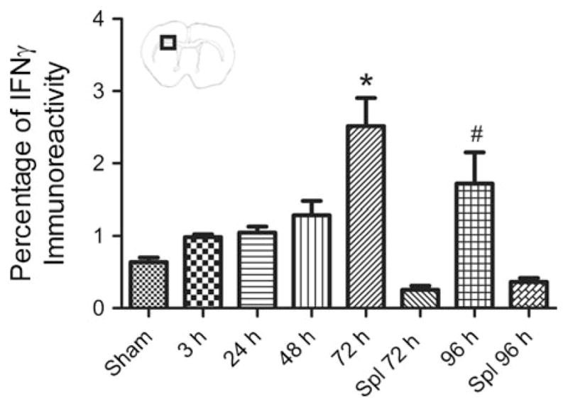
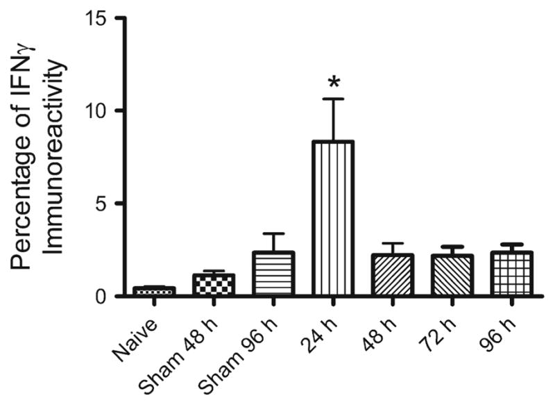
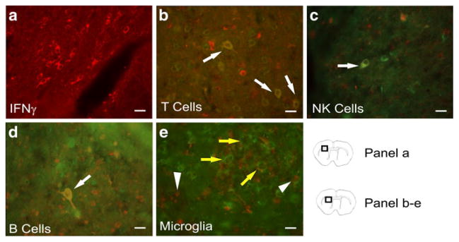
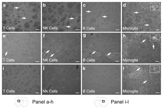
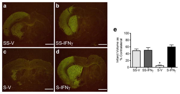
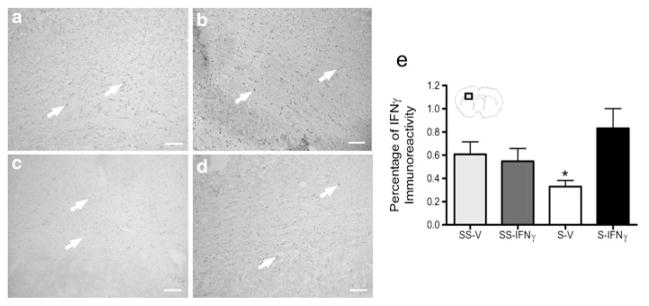
Delayed neuronal death associated with stroke has been increasingly linked to the immune response to the injury. Splenectomy prior to middle cerebral artery occlusion (MCAO) is neuroprotective and significantly reduces neuroinflammation. The present study investigated whether splenic signaling occurs through interferon gamma (IFNγ). IFNγ was elevated early in spleens but later in the brains of rats following MCAO. Splenectomy decreased the amount of IFNγ in the infarct post-MCAO. Systemic administration of recombinant IFNγ abolished the protective effects of splenectomy with a concurrent increase in INFγ expression in the brain. These results suggest a role for spleen-derived IFNγ in stroke pathology.


Conflict of interest statement

Conflicts of Interest

The authors have no conflicts of interest.

Figures

Fig. 1
Fig. 1
IFNγ levels increase in the injured brain post-MCAO. IFNγ immunohistochemistry of brain tissue from sham operated animals, animals that received splenectomies two weeks prior to MCAO and were euthanized at 72 and 96 h post-MCAO, and animals 3, 24, 48, 72, and 96 h post-MCAO. IFNγ protein levels were significantly higher at 72 and 96 h compared to sham operated animals and animals that received splenectomy prior to MCAO at 72 and 96 h post-MCAO (* p<0.01; # p<0.05). For each group n≥3. Box in brain graphic depicts area used for quantification of IFNγ levels. Sham denotes a sham MCAO and Spl denotes rats that underwent splenectomy prior to MCAO
Fig. 2
Fig. 2
Splenic IFNγ production is elevated at 24 h post-MCAO. Spleens from animals 24, 48, 72, 96 h post-MCAO along with naïve, 48 and 96 h sham-MCAO were assayed using immunohistochemistry for IFNγ. IFNγ protein levels were found to be significantly elevated 24 h post-MCAO (* p<0.0002) compared to the other groups
Fig. 3
Fig. 3
Representative brain sections from rats 96 h post-MCAO were stained with IFNγ and immune cell surface markers to identify what types of cells are expressing IFNγ in the infarct and peri-infarct. Micrographs show IFNγ (red) (a), and double staining merged images of IFNγ (red) with CD3 (green) for T cells (b), CD161 (green) for natural killer cells (c), and CD45R (green) for B cells (d); yellow cells with white arrows indicate areas of co-localization. A micrograph of staining with CD11b (green) for microglia/macrophages and IFNγ (red) (e) demonstrate a lack of co-localization of CD11b and IFNγ. In figure e, arrow heads indicate IFNγ positive cells and yellow arrows indicate CD11b positive cells. Scale bars=20 μm. Box in brain graphics depicts the regions where images were taken for a given micrograph
Fig. 4
Fig. 4
T cells, B cells, NK cells, and microglia/macrophages are present in the ipsilateral hemisphere following MCAO. At 96 h post-MCAO immunohistochemistry for immune cell surface markers shows peripheral immune cells are present in the ipsilateral hemisphere. Micrographs show CD3 positive cells (T cells) (a), CD161 positive cells (NK cells) (b), CD45R positive cells (B cells) (c), and CD11b positive cells (microglia/macrophages) (d) in the infarcted hemisphere. Micrographs from splenectomized rats demonstrate a decrease in immunostaining for T cells (e), NK cells (f), B cells (g), and microglia/macrophages (h) in the ipsilateral hemisphere. However in the contralateral hemisphere there is an absence of staining for T cells (i), NK cells (j), and B cells (k). Only microglia/macrophages were detected in the contralateral hemispheres (l). Inserts provide representative images of the morphological states of the microglia/macrophages present in each group and show an amoeboid cell (d), an amoeboid cell with evident ramifications (h), and a ramified cell (l). Scale bars=100 μm. The scale bar of the inserts=20 μm. Box in brain graphics depicts the regions where images were taken for a given micrograph
Fig. 5
Fig. 5
Recombinant IFNγ increases neural injury following MCAO in splenectomized rats. Recombinant IFNγ increases infarct volume in splenectomized rats at 96 h post-MCAO to levels not different from sham-splenectomized rats. Infarct volumes were measured as a percentage of the contralateral hemisphere with Fluoro-Jade staining. Graph depicts average infarct volumes for each group at 96 h post-MCAO (e). The splenectomy-vehicle treated rats had significantly lower infarcts than the other treatment groups (* p<0.0001). The splenectomy-IFNγ treated rats had infarcts that were not significantly different from the sham-splenectomy groups. Representative images for each treatment group at 96 h post-MCAO: sham-splenectomy-vehicle (SS-V) n=4 (a), sham-splenectomy-rIFNγ (SS-IFNγ) n=6 (b), splenectomy-vehicle (S-V) n= 4 (c), and splenectomy-rIFNγ (S-IFNγ) n=6 (d). Scale bars=2 mm
Fig. 6
Fig. 6
Recombinant IFNγ increases IFNγ expression in the infarct of splenectomized rats. The graph shows splenectomy results in a significant decrease in IFNγ protein expression at 96 h post-MCAO (* p< 0.02) (e). However rats that received splenectomy and rIFNγ had IFNγ protein levels not significantly different than the rats which underwent sham-splenectomy prior to MCAO. Representative images from each treatment group at 96 h following MCAO: sham-splenectomy-vehicle (SS-V) (a), sham-splenectomy-rIFNγ (SS-IFNγ) (b), splenectomy-vehicle (S-V) (c), and splenectomy-rIFNγ (S-IFNγ) (d). Box in brain graphics depicts the regions where images were taken for a given micrograph
Fig. 7
Fig. 7
Recombinant IFNγ is not cytotoxic to cultured primary neurons or OLs. Primary neuronal and OL cultures were treated with 20 ng/ml of rIFNγ under normoxic and OGD for 24 h. Recombinant IFNγ does not increase the amount of cell death, as measured by LDH, in neuronal cultures under normoxic or OGD conditions for 24 h (a). Oligodendrocytes subjected to 24 h of OGD and rIFNγ did not have significantly different survival rates (b). Representative images depict neuronal (c) and OL (d) cultures prior to treatment. Scale bars=30 μm

References

    1. Ajmo CT, Jr, Vernon DO, Collier L, Pennypacker KR, Cuevas J. Sigma receptor activation reduces infarct size at 24 hours after permanent middle cerebral artery occlusion in rats. Curr Neurovasc Res. 2006;3(2):89–98. - PubMed
    1. Ajmo CT, Jr, Vernon DO, Collier L, Hall AA, Garbuzova-Davis S, Willing A, Pennypacker KR. The spleen contributes to stroke-induced neurodegeneration. J Neurosci Res. 2008;86:2227–2234. - PMC - PubMed
    1. Bal-Price A, Brown GC. Inflammatory neurodegeneration mediated by nitric oxide from activated glia-inhibiting neuronal respiration, causing glutamate release and excitotoxicity. J Neurosci. 2001;21(17):6480–6491. - PMC - PubMed
    1. Barres BA, Schmid R, Sendnter M, Raff MC. Multiple extracellular signals are required for long-term oligodendrocyte survival. Development. 1993;118(1):283–295. - PubMed
    1. Becker KJ, Kindrick DL, Lester MP, Shea C, Ye ZC. Sensitization to brain antigens after stroke is augmented by lipopolysaccharide. J Cereb Blood Flow Metab. 2005;25(12):1634–1644. - PMC - PubMed

Publication types

MeSH terms