Review
. 2012 Jul 1;303(1):E1-17.
doi: 10.1152/ajpendo.00555.2011. Epub 2012 Feb 21.

cAMP signaling in skeletal muscle adaptation: hypertrophy, metabolism, and regeneration

Affiliations
Review

cAMP signaling in skeletal muscle adaptation: hypertrophy, metabolism, and regeneration

Rebecca Berdeaux et al. Am J Physiol Endocrinol Metab. .

Abstract

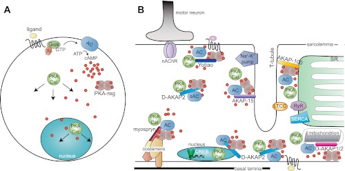
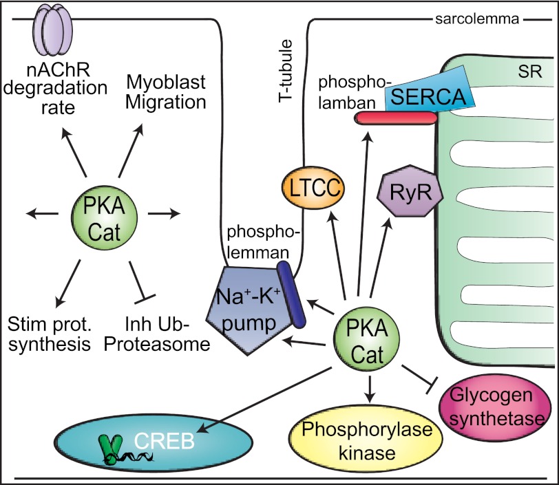
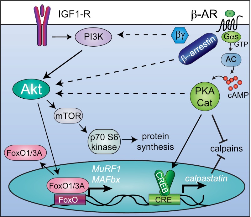
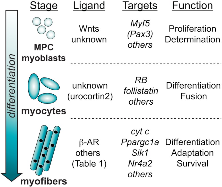
Among organ systems, skeletal muscle is perhaps the most structurally specialized. The remarkable subcellular architecture of this tissue allows it to empower movement with instructions from motor neurons. Despite this high degree of specialization, skeletal muscle also has intrinsic signaling mechanisms that allow adaptation to long-term changes in demand and regeneration after acute damage. The second messenger adenosine 3',5'-monophosphate (cAMP) not only elicits acute changes within myofibers during exercise but also contributes to myofiber size and metabolic phenotype in the long term. Strikingly, sustained activation of cAMP signaling leads to pronounced hypertrophic responses in skeletal myofibers through largely elusive molecular mechanisms. These pathways can promote hypertrophy and combat atrophy in animal models of disorders including muscular dystrophy, age-related atrophy, denervation injury, disuse atrophy, cancer cachexia, and sepsis. cAMP also participates in muscle development and regeneration mediated by muscle precursor cells; thus, downstream signaling pathways may potentially be harnessed to promote muscle regeneration in patients with acute damage or muscular dystrophy. In this review, we summarize studies implicating cAMP signaling in skeletal muscle adaptation. We also highlight ligands that induce cAMP signaling and downstream effectors that are promising pharmacological targets.


Figures

Fig. 1.
Fig. 1.
Models of cAMP signaling in muscle cells. A: classic view of cAMP production at the plasma membrane with diffusion into the cytoplasm. A ligand binds to a G protein-coupled receptor (GPCR), which activates Gαs. Gαs activates adenylyl cyclase (AC), which produces cAMP. cAMP binds to PKA regulatory subunits (PKA-reg), allowing dissociation of PKA catalytic subunits (PKA-cat), which diffuse into the cytoplasm and nucleus and phosphorylate target proteins. B: in a differentiated myofiber, cAMP production and PKA activity are localized to different subcellular compartments by anchoring proteins (AKAPs), including Yotiao (NMJ), AKAP-15 (T-tubules), AKAP-100 (SR), myospryn (costameres), D-AKAP2 (sarcoplasm, nucleus, sarcolemma, mitochondria), and D-AKAP1 (mitochondria). Protein complexes at the neuromuscular junction, on T-tubules, the SR, mitochondrion, and nucleus are shown. PKA-reg (in A, sienna) are not labeled in B. Several PKA substrates are shown including the Na+-K+ pump, L-type calcium channel (LTCC), and ryanodine receptor (RyR). PKA also modulates activity of the SERCA calcium pump and stability of nicotinic acetylcholine receptors (nAChR).
Fig. 2.
Fig. 2.
PKA targets in myofibers. Some specific substrates include the Na+-K+ pump, phospholemman, LTCC, RyR1, phospholamban (slow-twitch fibers only), glycogen synthetase, phosphorylase kinase, and CREB. Other general cellular processes for which specific targets are not clearly identified are also depicted: nAChR degradation, protein synthesis, the ubiquitin-proteasome pathway, and migration (undifferentiated myoblasts only). This is a partial depiction of PKA substrates.
Fig. 3.
Fig. 3.
GPCRs that induce myofiber hypertrophy. Four GPCRs (Fzd7, β2-AR, CRFR2, and LPA receptor) have been shown to stimulate hypertrophy in myotubes and/or myofibers. Cognate ligands are italicized. A partial view of the known signaling mediators is shown. CRFR2 stimulates myofiber growth by an uncharacterized effector pathway. β2-AR also induces fiber type transitions to fast-twitch fibers (not shown).
Fig. 4.
Fig. 4.
Possible mechanisms of Akt activation by β-AR signaling. IGF-I activates Akt, which stimulates protein synthesis via activating mTOR and inhibits muscle-specific ubiquitin ligase expression via repressive phosphorylation of FoxO transcription factors. β2-AR signaling leads to muscle hypertrophy, which is accompanied by activation of Akt, activation of protein synthesis, and inhibition of proteolysis. PKA signaling induces calpastatin transcription and inhibits calpains by an unknown mechanism. β2-AR signaling also activates Akt by an unknown mechanism, possibly mediated by Gβγ subunits, β-arrestin, or PKA (dashed arrows). Not shown: Wnt7a-Fzd7-Gαs activates PI 3-kinase directly.
Fig. 5.
Fig. 5.
Roles of CREB in muscle cells at different stages of differentiation. During development or after injury, myogenic precursor cells (MPCs) differentiate into myoblasts, myocytes, and myofibers. The known ligands that stimulate CREB, known CREB target genes, and CREB functions at each developmental stage are listed.

References

    1. Akimoto T, Pohnert SC, Li P, Zhang M, Gumbs C, Rosenberg PB, Williams RS, Yan Z. Exercise stimulates Pgc-1alpha transcription in skeletal muscle through activation of the p38 MAPK pathway. J Biol Chem 280: 19587– 19593, 2005 - PubMed
    1. Akimoto T, Sorg BS, Yan Z. Real-time imaging of peroxisome proliferator-activated receptor-γ coactivator-1α promoter activity in skeletal muscles of living mice. Am J Physiol Cell Physiol 287: C790– C796, 2004 - PubMed
    1. Ametller E, Busquets S, Fuster G, Figueras MT, De Oliveira CC, Toledo M, Korzeniewska K, Argiles JM, Lopez-Soriano FJ. Effects of formoterol on protein metabolism in myotubes during hyperthermia. Muscle Nerve 43: 268– 273, 2011 - PubMed
    1. Amieux PS, Howe DG, Knickerbocker H, Lee DC, Su T, Laszlo GS, Idzerda RL, McKnight GS. Increased basal cAMP-dependent protein kinase activity inhibits the formation of mesoderm-derived structures in the developing mouse embryo. J Biol Chem 277: 27294– 27304, 2002 - PubMed
    1. Antipenko A, Frias JA, Parra J, Cadefau JA, Cusso R. Effect of chronic electrostimulation of rabbit skeletal muscle on calmodulin level and protein kinase activity. Int J Biochem Cell Biol 31: 303– 310, 1999 - PubMed

Publication types