Review
. 2012 Mar;139(6):1059-69.
doi: 10.1242/dev.070730.

The hypoblast (visceral endoderm): an evo-devo perspective

Affiliations
Review

The hypoblast (visceral endoderm): an evo-devo perspective

Claudio D Stern et al. Development. 2012 Mar.

Abstract

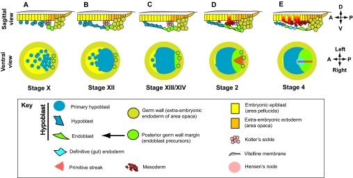
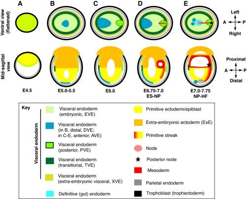
When amniotes appeared during evolution, embryos freed themselves from intracellular nutrition; development slowed, the mid-blastula transition was lost and maternal components became less important for polarity. Extra-embryonic tissues emerged to provide nutrition and other innovations. One such tissue, the hypoblast (visceral endoderm in mouse), acquired a role in fixing the body plan: it controls epiblast cell movements leading to primitive streak formation, generating bilateral symmetry. It also transiently induces expression of pre-neural markers in the epiblast, which also contributes to delay streak formation. After gastrulation, the hypoblast might protect prospective forebrain cells from caudalizing signals. These functions separate mesendodermal and neuroectodermal domains by protecting cells against being caught up in the movements of gastrulation.


Figures

Fig. 1.
Fig. 1.
Development of the chick hypoblast and endoderm. Top row: sections through the posterior part of the embryo; posterior (P) to the right, dorsal (D; epiblast) towards the top. Bottom row: ventral views of the whole embryo showing main anatomical features; posterior to the right, left side towards the top. Pre-primitive streak stages are shown in Roman numbers according to Eyal-Giladi and Kochav (Eyal-Giladi and Kochav, 1976). Primitive streak and later stages are shown in Arabic numerals according to Hamburger and Hamilton (Hamburger and Hamilton, 1951). (A) Stage X (freshly laid embryo). The ventral surface is peppered with islands of primary hypoblast. Peripherally, there is a multilayered extra-embryonic endoderm (germ wall). Koller’s sickle (pink) marks the posterior border of the area pellucida. (B) Stage XII. The islands of primary hypoblast have merged into a layer that covers the posterior half of the embryo. (C) Stage XIII/XIV. The primary hypoblast now makes a complete layer covering almost the entire area pellucida; the first (sickle) endoblast cells (green) appear posteriorly, derived from the posterior germ wall margin (green circles). (D) Stage 2. The endoblast continues to displace the primary hypoblast anteriorly. The primitive streak has appeared and cells now delaminate from the posterior epiblast to form mesoderm. (E) Stage 4. The primitive streak has now elongated to about two-thirds of the diameter of the area pellucida. Definitive (future gut) endoderm (light blue) has now inserted into the endodermal layer.
Fig. 2.
Fig. 2.
The visceral endoderm (VE) during early mouse development (∼E4.5-7.75, pre-streak to head fold stages). Top row: views from the visceral endodermal side (parietal endoderm and associated trophoblast, which would obliterate this view, are shown only on the periphery of each figure); posterior (P) to the right, left side to the top. Bottom row: sagittal sections through the conceptus; posterior to the right, maternal (implantation or proximal) side is towards the top. (A) E4.5. The implanting blastocyst contains epiblast (yellow), VE (green) and parietal endoderm (gray). Trophoblast (black) is indicated. (B) At ∼E5.0-5.5, the distal visceral endoderm (DVE) forms a group of cells within the embryonic visceral endoderm (EVE). (C) By ∼E6.0, the DVE has spread to the anterior embryonic/extra-embryonic border, where it is now called anterior visceral endoderm (AVE). At around the same time, a molecularly distinct posterior visceral endoderm (PVE) emerges. (D) E6.75-7.0 [early streak (ES)-neural plate (NP) stage (Downs and Davies, 1993)]. By ∼E6.75, the primitive streak has appeared in the posterior axial midline of the epiblast, and extends anteriorly. (E) By E7.0 [NP-headfold (HF) stages], the anterior end of the primitive streak has condensed into the node (pink) and posteriorly it extends into the extra-embryonic region where, perhaps together with transitional visceral endoderm (TVE) and PVE, it condenses into the ‘allantoic core domain’ (ACD) or posterior node (Downs et al., 2009). By this time, the AVE seems to disappear into the anterior extra-embryonic visceral endoderm (XVE), though some AVE cells (dark blue) might remain interspersed with definitive (gut) endoderm (Kwon et al., 2008). With the exception of the DVE, which is morphologically distinct from the EVE, the EVE is characterized by its flat squamous cells, largely without microvilli or apical vacuoles. TVE is cuboidal containing some microvilli and vacuoles, and XVE is columnar with many microvilli and apical vacuoles (Bonnevie, 1950; Enders et al., 1978; Hogan and Tilly, 1981; Solter et al., 1970).

Similar articles

Cited by

References

    1. Acampora D., Di Giovannantonio L. G., Di Salvio M., Mancuso P., Simeone A. (2009). Selective inactivation of Otx2 mRNA isoforms reveals isoform-specific requirement for visceral endoderm anteriorization and head morphogenesis and highlights cell diversity in the visceral endoderm. Mech. Dev. 126, 882–897 - PubMed
    1. Albazerchi A., Stern C. D. (2007). A role for the hypoblast (AVE) in the initiation of neural induction, independent of its ability to position the primitive streak. Dev. Biol. 301, 489–503 - PubMed
    1. Albazerchi A., Cinquin O., Stern C. D. (2007). A new method to transfect the hypoblast of the chick embryo reveals conservation of the regulation of an Otx2 enhancer between mouse and chick extraembryonic endoderm. BMC Dev. Biol. 7, 25 - PMC - PubMed
    1. Arendt D., Nübler-Jung K. (1999). Rearranging gastrulation in the name of yolk: evolution of gastrulation in yolk-rich amniote eggs. Mech. Dev. 81, 3–22 - PubMed
    1. Artus J., Piliszek A., Hadjantonakis A. K. (2011). The primitive endoderm lineage of the mouse blastocyst: sequential transcription factor activation and regulation of differentiation by Sox17. Dev. Biol. 350, 393–404 - PMC - PubMed

Publication types