. 2012 Apr;241(4):663-74.
doi: 10.1002/dvdy.23755. Epub 2012 Feb 21.

Asymmetric requirement of surface epithelial β-catenin during the upper and lower jaw development

Affiliations
Free PMC article

Asymmetric requirement of surface epithelial β-catenin during the upper and lower jaw development

Ye Sun et al. Dev Dyn. 2012 Apr.
Free PMC article

Abstract

Background: Intercellular communication between epithelial and mesenchymal cells is central to mammalian craniofacial development. β-catenin is the gateway of canonical Wnt signaling, one of the major evolutionarily conserved cell-cell communication pathways in metazoa. In this study, we report an unexpected stage- and tissue-specific function of β-catenin during mammalian jaw development.

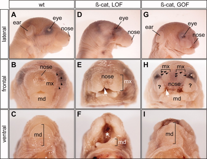
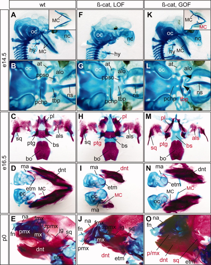
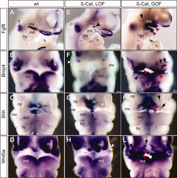
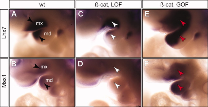
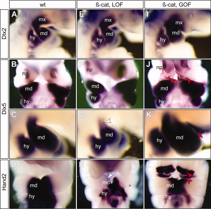
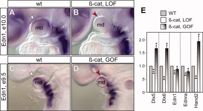
Results: Using a unique mouse genetic tool, we have discovered that epithelial β-catenin is essential for lower jaw formation, while attenuation of β-catenin is required for proper upper jaw development. Changes in β-catenin in vivo alter major epithelial Fgf8, Bmp4, Shh, and Edn1 signals, resulting in partial transcriptional reprogramming of the neural crest-derived mesenchyme, the primary source of jawbones.

Conclusions: The Wnt/β-catenin signal coordinates expression of multiple epithelial signals and has stage-specific asymmetric functions during mammalian upper and lower jaw development. In addition, these findings suggest that evolutionary changes of the canonical Wnt/β-catenin signaling pathway may lead to innovation of jaws.


Figures

Fig. 1
Fig. 1
Epithelial-specific Pitx1-Cre transgenic mice. X-gal staining (blue) of Cre activities of the Pitx1/Cre and R26RlacZ double heterozygous embryos. A–C: Whole-mount staining. D,E: Middle sagittal sections. Arrowheads point to the caudal limits of Cre activity; asterisk, ventral nasal placode; fnp, frontal nasal prominence; lnp, lateral nasal prominence; md, mandible prominence; mnp, medial nasal prominence; mx, madillary prominence; p, pituitary; pl, palate; t, tongue.
Fig. 2
Fig. 2
Conditional manipulation of epithelial β-catenin. A–C: Immunohistochemical analyses of sagittal cryostat sections from E10.5 wild-type (A, wt) and β-catenin loss-of-function (B, LOF) and gain-of-function (C, GOF) mutants using β-catenin-specific antibody. bracket, epithelial subdomain where epithelial β-catenin is conditionally altered; md, mandibular; RP, Rathke's pouch.
Fig. 3
Fig. 3
Asymetric requirements for epithelial β-catenin in upper and lower jaw formation. D–F: Newborn lower jaw defects of epithelial-specific β-catenin loss-of-function mutants (β-cat, LOF), and upper jaw defects of the gain-of-function mutants (β-cat, GOF, G-I). A–C: Wild-type controls. White arrow, cleft lips; black arrow, whiskers; asterisk, cleft palate; md, mandible; mx, maxillary; question mark, ectopic soft tissue.
Fig. 4
Fig. 4
Cartilage and bone defects of epithelial-specific β-catenin mutants. A–O: Alcian blue cartilage staing of of embryonic day (E) 14.5 (A,B,F,G,K,L), E16.5 embryos (C,D,H,I,M,N), and newborn pups (E,J,O) and alizarin red bone staining of E16.5 embryos and newborn pups. A–E: Wild-type controls. F–J: β-cateLOF mutants display severe truncation of Meckel's cartilage (MC) and dentary bone, deformation of palatine (pl) and pterygoid (ptg). K–O: β-cateGOF mutants have ectopic MC and upper jaw defects. Red labels indicate defective structures. All pictures are ventral views except A, E, F, J, K and O, which are sagittal views. Inserts in A and K show ventral view of dissected MC. Note that the dentary bones of the β-cateGOF (N) are connected distally but dissociated during staining and dissection process. Arrow, gap; arrowhead, unidentified cartilage rod; at, ala temporalis; alo, ala orbitalis; als, alisphenoid; bo, basioccipital; bs, basisphenoid; dnt, dentary; etm, ectotympanic; fmx, frontal process of maxilla; fn, frontal nasal cartilage; hy, hyoid; jg, jugal; ma, malleus; mx, maxilla; mc, Meckel's cartilage; na, nasal bone; nc, nasal capsule; ns, nasal septum; oc, otic capsule; pchp, parachordal plate; pl, palatine; ppso, pila postoptica; ptg, pterygoid; sq, squamosal; tbp, trabecular basal plate; zpmx, zygomatic process of maxilla.
Fig. 5
Fig. 5
β-catenin functions upstream of other epithelial signals. E–L: RNA in situ hybridization (purple color) of embryonic day (E) 10.5 embryos using gene-specific probes indicates decreased expression of Fgf8, Bmp4, Shh, and Wnt5a in the β-catenin loss-of-function (β-cat, LOF) mutants (E–H), while expression of these genes were enhanced in the β-catenin gain-of-function (β-cat, GOF) mutants (I–L). A–D: Wild-type controls. I–L: Patchy ectopic staining (black arrows) of Fgf8 (I), Bmp4 (J), Shh (K), and Wnt5a (L) were observed in the β-catenin GOF mutants. All pictures are frontal views except A, E and I, which are lateral views. di, diencephalon; FEZ, frontonasal ectoderm zone; h, heart; hy, hyoid arch; md, mandibular prominence; mx, maxillary prominence; n, notochord/floor plate; np, nasal placode; o, otic vesicle; te, telencephalon; black arrowhead, residual expression of Fgf8 at the cleft of between mx and md; white arrowhead, reduced expression; red arrowhead, enhanced expression.
Fig. 6
Fig. 6
Epithelial β-catenin indirectly regulates expression of genes in the mesenchymal cells. Whole-mount RNA in situ hybridization using Lhx7 and Msx1 probes. A,B: Wild-type controls. C,D: Epithelial-specific β-catenin loss-of-function (β-cat, LOF) mutants. E,F: β-catenin gain-of-function (β-cat, GOF) mutants. black arrowhead, normal expression pattern; white arrowhead, reduced expression; red arrowhead, increased expression.
Fig. 7
Fig. 7
A–L: β-catenin induces partial reprogramming of maxillary mesenchyme. While expression of Dlx2 is maintained in both β-catenin loss-of-function (β-cat, LOF) mutants (E) and gain-of-function (β-cat, GOF) mutants (I), expression of the mdPA1 genes Dlx5 (B,C,F,G,J,K) and Hand2 (D,H,L) are reduced in the LOF mutants (white arrowheads in G and H), and ectopically induced in the GOF mutants (red arrowheads in K and L). A,E,I,C,G,K: Lateral views and the rest are frontal views. See Figure 4 for abbreviations.
Fig. 8
Fig. 8
Expression of Edn1 depends on epithelial β-catenin. A–D: Whole-mount RNA in situ hybridization using Edn1 specific probe. white arrowhead, normal expression in mandibular prominence (md); red arrowhead, reduced expression in mutants. expression in hyoid arch (hy) and the rest of caudal arches are unaffected. E: Real-time quantitative polymerase chain reaction analyses of relative expression levels of genes (x-axis) in the microdissected first pharyngeal arch at embryonic day (E) 10.5 using β-actin as an internal control. Asterisk, P < 0.05 (y-axis), Student t-test, n = 4.

Similar articles

Cited by

References

    1. Abzhanov A, Protas M, Grant BR, Grant PR, Tabin CJ. Bmp4 and morphological variation of beaks in Darwin's finches. Science. 2004;305:1462–1465. - PubMed
    1. Beverdam A, Merlo GR, Paleari L, Mantero S, Genova F, Barbieri O, Janvier P, Levi G. Jaw transformation with gain of symmetry after Dlx5/Dlx6 inactivation: mirror of the past? Genesis. 2002;34:221–227. - PubMed
    1. Brault V, Moore R, Kutsch S, Ishibashi M, Rowitch DH, McMahon AP, Sommer L, Boussadia O, Kemler R. Inactivation of the beta-catenin gene by Wnt1-Cre-mediated deletion results in dramatic brain malformation and failure of craniofacial development. Development. 2001;128:1253–1264. - PubMed
    1. Brito JM, Teillet MA, Le Douarin NM. An early role for sonic hedgehog from foregut endoderm in jaw development: ensuring neural crest cell survival. Proc Natl Acad Sci U S A. 2006;103:11607–11612. - PMC - PubMed
    1. Brito JM, Teillet MA, Le Douarin NM. Induction of mirror-image supernumerary jaws in chicken mandibular mesenchyme by Sonic Hedgehog-producing cells. Development. 2008;135:2311–2319. - PubMed

Publication types

LinkOut - more resources