. 2012 May 1;94(2):379-90.
doi: 10.1093/cvr/cvs096. Epub 2012 Feb 21.

Loss of the miR-144/451 cluster impairs ischaemic preconditioning-mediated cardioprotection by targeting Rac-1

Affiliations

Loss of the miR-144/451 cluster impairs ischaemic preconditioning-mediated cardioprotection by targeting Rac-1

Xiaohong Wang et al. Cardiovasc Res. .

Abstract

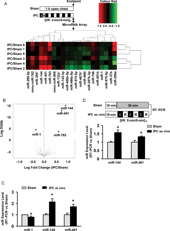
Aims: While a wealth of data has uncovered distinct microRNA (miR) expression alterations in hypertrophic and ischaemic/reperfused (I/R) hearts, little is known about miR regulation and response to ischaemic preconditioning (IPC).

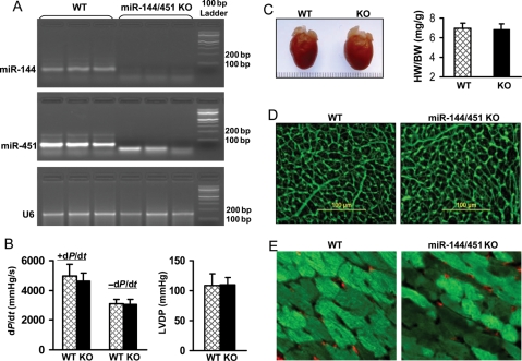
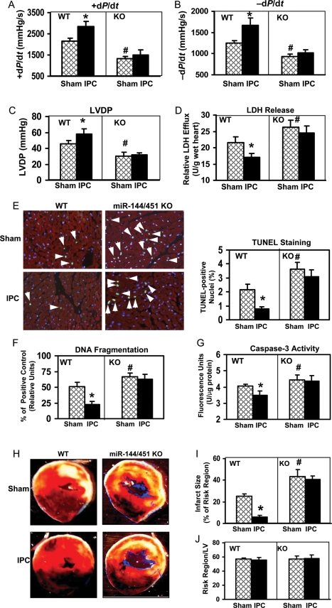
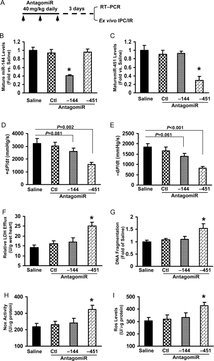
Methods and results: We analysed miRs in murine hearts preconditioned with six cycles of 4 min ischaemia via coronary artery occlusion, followed by 4 min reperfusion in vivo. Both miRs within the miR-144/451 cluster were the most elevated among a cohort of 21 dysregulated miRs in preconditioned hearts, compared with shams. To investigate the significance of this finding, we examined IPC-mediated cardioprotection within a miR-144/451-knockout (KO) mouse model. Wild-type (WT) hearts exposed to IPC followed by I/R (30 min/24 h) showed a smaller infarction size compared with mice treated with I/R alone. In contrast, IPC failed to protect miR-144/451-KO hearts against infarct caused by I/R treatment. Thus, the miR-144/451 cluster is required for IPC-elicited cardioprotection. Rac-1, a key component of NADPH oxidase, was mostly up-regulated in KO hearts among three bona fide targets (Rac-1, 14-3-3ζ, and CUGBP2) for both miR-144 and miR-451. Accordingly, reactive oxygen species (ROS) levels were markedly increased in KO hearts upon IPC, compared with IPC-WT hearts. Pre-treatment of KO hearts with a Rac-1 inhibitor NSC23766 (20 mg/kg, ip) reduced IPC-triggered ROS levels and restored IPC-elicited cardioprotection. Using antagomiRs, we showed that miR-451 was largely responsible for IPC-mediated cardioprotection.

Conclusion: Loss of the miR-144/451 cluster limits IPC cardioprotection by up-regulating Rac-1-mediated oxidative stress signalling.


Figures

Figure 1
Figure 1
miRNA expression profile in IPC hearts. (A) A heat-map of the up-regulated and down-regulated miRNAs in murine hearts subjected to six cycles of 4 min LAD occlusion and 4 min reperfusion, compared with sham groups. All of the miR array raw data are available in Supplementary material online, Table S1. (B) Microarray data are summarized by a volcano plot graph, which displays both fold-change and t-test criteria (log odds). miR-1, -144, -451, and -762 are the most significantly dysregulated miRs in IPC hearts compared with shams. (C) Alterations in expression levels of miR-1, -144, and -451 were validated by qRT–PCR (normalized to control U6). (D) Both miR-144 and miR-451 were significantly up-regulated in murine hearts upon ex vivo IPC (n = 6, *P< 0.05).
Figure 2
Figure 2
Characterization of miR-144/451-deficient hearts. (A) Stem-loop RT–PCR indicated that both miR-144 and miR-451 expression were absent in miR-144/451-KO hearts. (B) Rates of myocardial contraction (+dP/dt) and relaxation (–dP/dt), (C) the ratio of heart weight/body weight (HW/BW), (D) cardiomyocyte size stained with Oregon Green 488-labelled wheat germ agglutinin, and (E) the density of blood vessel stained with CD31 (red) were shown to be similar to WT hearts (n = 6 for measurement of function, n = 10 for determining the ratio of HW/BW, and n = 4 for immunostaining).
Figure 3
Figure 3
Ablation of the miR-144/451 cluster impairs IPC-induced efficacy on post-I/R functional recovery and myocardial damage. (A) Rates of myocardial contraction (+dP/dt) and (B) relaxation (–dP/dt), and (C) LVDP all showed better recovery in WT hearts upon IPC, whereas these measures were impaired in miR-144/451-null hearts, compared with shams. (D) Total LDH in coronary effluent, collected during the first 10 min of reperfusion, was significantly decreased in IPC-WT hearts compared with the sham group, whereas no difference was observed in miR-144/451-KO hearts between IPCs and shams. (*P < 0.05 vs. WT sham; #P < 0.05 vs. WT sham, n = 11 for AD). (E) Triple-staining with anti-α-sarcomeric actin antibody (red), DAPI (blue), and TUNEL (green) to determine apoptosis and quantification in miR-144/451-null hearts upon IPC and sham treatment (n = 5, with three sections from each heart). (F) Histone-associated DNA fragmentation was determined by a cell-death-detection ELISA kit (Roche Applied Science). (G) Caspase-3 activity was measured by Caspase-3/CPP32 Fluorometric Assay kit (BioVision). *P < 0.05 vs. WT sham; #P < 0.05 vs. WT sham, n = 5 for (F) and (G). Myocardial infarct size was greatly reduced in WT hearts upon in vivo IPC with six cycles of 4 min LAD occlusion/4 min reperfusion followed by I/R (30 min/24 h) compared with the sham I/Rs, whereas there was no difference in miR-144/451-null hearts subjected to either IPC-I/R or sham-I/R (H and I). (J) Region at risk was not significantly different between groups. *P < 0.05 vs. WT sham; #P < 0.05 vs. WT sham, n = 9.
Figure 4
Figure 4
Ablation of miR-144/451 activates the Rac-1-mediated oxidative stress signalling pathway. (A) 14-3-3ζ, Rac-1, and CUGBP2 are confirmed targets for both miR-144 and miR-451, and previous studies indicate that 14-3-3ζ and CUGBP2 act upstream of Rac-1. (B) Rac-1, but not 14-3-3ζ and CUGBP2, was significantly up-regulated in miR-144/451-null hearts compared with WTs. *P < 0.05 vs. WT, n = 6. (C) Protein levels of Rac-1 were significantly reduced in mouse hearts upon in vivo and ex vivo IPC, relative to shams. *P < 0.05 vs. sham, n = 6. (D) The activity of NADPH oxidase (Nox) and (E) the ROS levels were dramatically increased in IPC-miR-144/451-null hearts, compared with IPC-WTs, suggesting excessive oxidative stress, leading to elevation of LDH release (F). *P < 0.05 vs. sham; #P < 0.05 vs. IPC-WT, n = 5.
Figure 5
Figure 5
Pre-treatment of miR-144/451-null mice with an Rac-1 inhibitor NSC23766 restores IPC-induced cardioprotection. (A) The experiment protocol. (B) The Nox activity and (C) ROS levels were significantly reduced in miR-144/451-null hearts treated with NSC23766, compared with the saline controls. (D and E) NSC23766-treated miR-144/451-null hearts displayed an improvement in the post-I/R functional recovery (±dP/dt), compared with those given saline treatment. (FH) Myocardial I/R-induced cellular damage (F, LDH release; G, DNA fragmentation; H, caspase-3 activity) was attenuated in miR-144/451-null hearts upon pre-inhibition of Rac-1 by NSC23766, compared with saline controls. *P < 0.05 vs. saline, n = 6.
Figure 6
Figure 6
miR-451 plays a dominant role in the miR-144/451-mediated action. (A) A protocol for antagomiR treatment: WT mice (B6129SF2/J F2, 6 weeks old, male) received either antagomiR-144, antagomiR-451, or antagomiR control, or a comparable volume of saline (200 mL) through three consecutive daily tail vein injections (3 × 40 mg/kg body weight). Ex vivo IPC-I/R was performed at the third day after the final injection. (B and C) Stem-loop RT–PCR determined miR-144 or miR-451 expression in antagomiR-treated hearts. n = 4, *P < 0.001 vs. saline- and mutant-treated controls. (D and E) IPC-induced myocardial function recovery was impaired in antagomiR-451- but not in antagomiR-144-treated hearts; accordingly (F and G), myocardial injury (necrosis: LDH release and apoptosis: DNA fragmentation) was increased in antagomiR-451- but not in antagomiR-144-treated hearts. (H and I) Oxidative stress (Nox activity and ROS levels) was significantly activated in antagomiR-451- but not in antagomiR-144-treated hearts. *P < 0.05 vs. antagomiR control, n = 6.

Similar articles

Cited by

References

    1. Yellon DM, Downey JM. Preconditioning the myocardium: from cellular physiology to clinical cardiology. Physiol Rev. 2003;83:1113–1151. - PubMed
    1. Otani H. Ischemic preconditioning: from molecular mechanisms to therapeutic opportunities. Antioxid Redox Signal. 2008;10:207–247. - PubMed
    1. Budas GR, Jovanovic S, Crawford RM, Jovanovic A. Hypoxia-induced preconditioning in adult stimulated cardiomyocytes is mediated by the opening and trafficking of sarcolemmal KATP channels. FASEB J. 2004;18:1046–1058. - PMC - PubMed
    1. Budas GR, Sukhodub A, Alessi DR, Jovanović A. 3′Phosphoinositide-dependent kinase-1 is essential for ischemic preconditioning of the myocardium. FASEB J. 2006;20:2556–2568. - PMC - PubMed
    1. Sukhodub A, Jovanović S, Du Q, Budas G, Clelland AK, Shen M, et al. AMP-activated protein kinase mediates preconditioning in cardiomyocytes by regulating activity and trafficking of sarcolemmal ATP-sensitive K(+) channels. J Cell Physiol. 2007;210:224–236. - PMC - PubMed

Publication types

MeSH terms