Comparative Study
. 2012 Mar 1;3(1):e00318-11.
doi: 10.1128/mBio.00318-11. Print 2012.

Comparative genomics of enterococci: variation in Enterococcus faecalis, clade structure in E. faecium, and defining characteristics of E. gallinarum and E. casseliflavus

Affiliations
Comparative Study

Comparative genomics of enterococci: variation in Enterococcus faecalis, clade structure in E. faecium, and defining characteristics of E. gallinarum and E. casseliflavus

Kelli L Palmer et al. mBio. .

Abstract

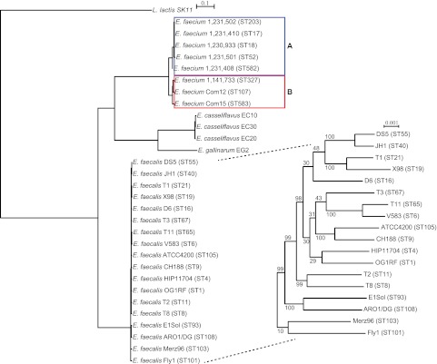
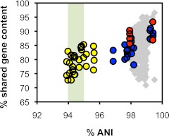
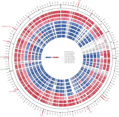
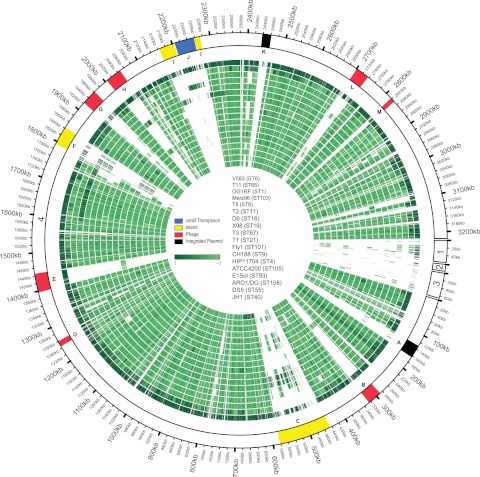
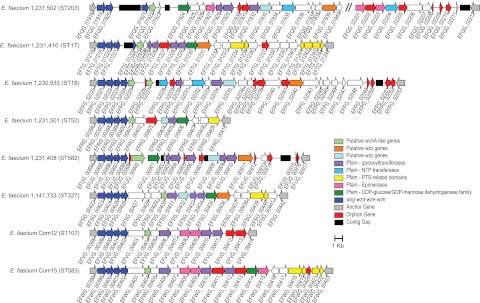
The enterococci are Gram-positive lactic acid bacteria that inhabit the gastrointestinal tracts of diverse hosts. However, Enterococcus faecium and E. faecalis have emerged as leading causes of multidrug-resistant hospital-acquired infections. The mechanism by which a well-adapted commensal evolved into a hospital pathogen is poorly understood. In this study, we examined high-quality draft genome data for evidence of key events in the evolution of the leading causes of enterococcal infections, including E. faecalis, E. faecium, E. casseliflavus, and E. gallinarum. We characterized two clades within what is currently classified as E. faecium and identified traits characteristic of each, including variation in operons for cell wall carbohydrate and putative capsule biosynthesis. We examined the extent of recombination between the two E. faecium clades and identified two strains with mosaic genomes. We determined the underlying genetics for the defining characteristics of the motile enterococci E. casseliflavus and E. gallinarum. Further, we identified species-specific traits that could be used to advance the detection of medically relevant enterococci and their identification to the species level.


Figures

FIG 1
FIG 1
Core gene tree. Concatenated sequences of 847 genes core to 30 enterococci and the outgroup species Llactis were aligned, and a phylogenetic tree was generated using RAxML with bootstrapping. The bootstrap value for all nodes outside the Efaecalis clade is 100. Efaecium clades A (blue) and B (red) are indicated.
FIG 2
FIG 2
ANI plot. Each point represents a pairwise comparison of two genomes. Grey diamonds, Efaecalis-Efaecalis comparisons. Blue circles, clade A E. faecium-clade A E. faecium comparisons. Red circles, clade B E. faecium-clade B E. faecium comparisons. Yellow circles, clade A E. faecium-clade B E. faecium comparisons. A species threshold of 94 to 95% ANI is indicated by the green-shaded area.
FIG 3
FIG 3
Efaecium genome mosaicism plot. The outermost ring shows Efaecium Com12 scaffolds, ordered by decreasing length clockwise from scaffold 1, with each gene represented as a radial position along the ring. Each of the remaining 7 Efaecium genomes is represented by the rings below Com12. Genes are colored by membership in clade A (blue) or clade B (red), as determined by individual gene trees built from ortholog groups. The strains shown, from the outermost to the innermost rings, are Com12, 733, Com15, 501, 408, 502, 933, and 410. The locations of dnaA, Com12 MLST alleles, pbp5, and the EfmCRISPR1-cas locus are shown.
FIG 4
FIG 4
Efaecalis genome mosaicism plot. The outermost ring shows Efaecalis V583 chromosomal (scaffold 4) and plasmid scaffolds (scaffold 1, pTEF2; scaffold 2, pTEF3; scaffold 3, pTEF1), with each gene represented as a radial position along the ring. Each of the remaining 17 Efaecalis genomes is represented by the rings below V583. Genes are colored by phylogenetic distance from Efaecalis V583 (from dark to light green with increasing phylogenetic distance), as determined by individual gene trees built from ortholog groups. The strains shown, from the outermost to the innermost rings, are V583, T11, OG1RF, Merz96, T8, T2, D6, X98, T3, T1, Fly1, CH188, HIP11704, ATCC 4200, E1Sol, AR01/DG, DS5, and JH1. The locations of Efaecalis variable regions are shown (9). A, integrated plasmid; B, prophage 1; C, Efaecalis pathogenicity island; D, prophage 2; E, prophage 3; F, putative island; G, prophage 4; H, prophage 5; I, putative island; J, vancomycin resistance (vanB) transposon; K, integrated plasmid; L, prophage 6; M, prophage 7.
FIG 5
FIG 5
Putative Efaecium capsule loci. The core wzg-wzd-wze-wzh genes and downstream variable region are shown for 8 Efaecium strains. Conserved anchor genes flanking the core and variable regions are indicated. Variable region genes are colored by BLASTP and Pfam conserved-domain hits shown in data set S4 in the supplemental material. Multiple Pfam domains were collapsed into categories (for example, glycosyltransferases). Only the most abundant Pfam categories are shown. Orphan genes not grouped by OrthoMCL are indicated. Contig gaps in scaffolds are indicated by black bars; the size of the black bar is proportional to the number of N’s inserted during genome assembly. In Efaecium 502, the nucleotide sequence of wze is conserved but is interrupted by a contig gap, and a scaffold gap occurs between wzh and the EFVG_00414 flanking gene homologue (indicated by vertical slashes). The drawing is to scale, and a scale bar is shown.

Similar articles

Cited by

References

    1. Aarestrup FM, Butaye P, Witte W. 2002. Nonhuman reservoirs of enterococci, p 55–99 In Gilmore MS, The enterococci: pathogenesis, molecular biology, and antibiotic resistance. ASM Press, Washington, DC
    1. Malani PN, Kauffman CA, Zervos MJ. 2002. Enterococcal disease, epidemiology, and treatment, p 385–408 In Gilmore MS, The enterococci: pathogenesis, molecular biology, and antibiotic resistance. ASM Press, Washington, DC
    1. Tannock GW, Cook G. 2002. Enterococci as members of the intestinal microflora of humans, p 101–132 In Gilmore MS, The enterococci: pathogenesis, molecular biology, and antibiotic resistance. ASM Press, Washington, DC
    1. Willems RJ, Hanage WP, Bessen DE, Feil EJ. 2011. Population biology of gram-positive pathogens: high-risk clones for dissemination of antibiotic resistance. FEMS Microbiol. Rev. 35:872–900 - PMC - PubMed
    1. Paulsen IT, et al. 2003. Role of mobile DNA in the evolution of vancomycin-resistant Enterococcus faecalis. Science 299:2071–2074 - PubMed

Publication types

Substances

LinkOut - more resources