. 2012 Apr 6;287(15):12353-64.
doi: 10.1074/jbc.M112.340893. Epub 2012 Feb 21.

Susceptibility of β1 Na+-K+ pump subunit to glutathionylation and oxidative inhibition depends on conformational state of pump

Affiliations

Susceptibility of β1 Na+-K+ pump subunit to glutathionylation and oxidative inhibition depends on conformational state of pump

Chia-Chi Liu et al. J Biol Chem. .

Abstract

Glutathionylation of cysteine 46 of the β1 subunit of the Na(+)-K(+) pump causes pump inhibition. However, the crystal structure, known in a state analogous to an E2·2K(+)·P(i) configuration, indicates that the side chain of cysteine 46 is exposed to the lipid bulk phase of the membrane and not expected to be accessible to the cytosolic glutathione. We have examined whether glutathionylation depends on the conformational changes in the Na(+)-K(+) pump cycle as described by the Albers-Post scheme. We measured β1 subunit glutathionylation and function of Na(+)-K(+)-ATPase in membrane fragments and in ventricular myocytes. Signals for glutathionylation in Na(+)-K(+)-ATPase-enriched membrane fragments suspended in solutions that preferentially induce E1ATP and E1Na(3) conformations were much larger than signals in solutions that induce the E2 conformation. Ouabain further reduced glutathionylation in E2 and eliminated an increase seen with exposure to the oxidant peroxynitrite (ONOO(-)). Inhibition of Na(+)-K(+)-ATPase activity after exposure to ONOO(-) was greater when the enzyme had been in the E1Na(3) than the E2 conformation. We exposed myocytes to different extracellular K(+) concentrations to vary the membrane potential and hence voltage-dependent conformational poise. K(+) concentrations expected to shift the poise toward E2 species reduced glutathionylation, and ouabain eliminated a ONOO(-)-induced increase. Angiotensin II-induced NADPH oxidase-dependent Na(+)-K(+) pump inhibition was eliminated by conditions expected to shift the poise toward the E2 species. We conclude that susceptibility of the β1 subunit to glutathionylation depends on the conformational poise of the Na(+)-K(+) pump.


Figures

FIGURE 1.
FIGURE 1.
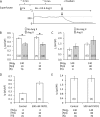
Conformation dependence of β1 subunit glutathionylation of pig kidney Na+-K+-ATPase. A, glutathionylation after incubation of Na+-K+-ATPase-enriched membrane fragments in solution containing 0.1 mm EDTA, 25 mm histidine, and 2 mm Na2ATP at a pH of 7.1 (E1ATP) or in solution containing 0.1 mm EDTA and 25 mm histidine at a pH of 7.1 (E2). Glutathionylation was measured with the biotin-GSH technique. B, glutathionylation in E1ATP and E2 conformations with and without 10 mm KCl in 30 mm histidine solution. Glutathionylation was measured with the GSH antibody technique. C, glutathionylation after incubation of Na+-K+-ATPase in 100 mm NaCl, 4 mm MgCl2, and 20 mm histidine at a pH of 7.35 (E1Na3). The solution used to promote the E2 conformation contained 20 mm histidine at a pH of 7.35 in these experiments. Glutathionylation was measured with the GSH antibody technique. D, glutathionylation induced by ONOO (ONOO) in E1ATP and E2 conformations. The solution used to promote the E2 conformation contained 0.1 mm EDTA and 25 mm histidine at a pH of 7.1 in these experiments. Glutathionylation was measured with the biotin-GSH technique. E, effect of pre-exposure to ouabain before exposure to ONOO on glutathionylation in the E2 conformation (0.1 mm EDTA, 25 mm histidine, pH of 7.1). F, glutathionylation with pre-exposure to ONOO before exposure to ouabain. The histograms show mean densitometry ± S.E. of five experiments. *, p < 0.05. IB, immunoblot; IP, immunoprecipitation.
FIGURE 2.
FIGURE 2.
A, SDS-PAGE of trypsin-treated pig Na+-K+-ATPase-enriched membrane fragments. The E2 conformation was stabilized by incubation in 30 mm histidine at a pH of 7.4 alone. The E1Na3 conformation (E1) was stabilized by the further addition of 4 mm MgCl2 and 100 mm NaCl. The Na+-K+-ATPase was exposed to 0.5 mm ONOO and 0.5 mm GSH before exposure to trypsin for 20 min as indicated and compared with controls. Controls received 5 μg of trypsin/100 μg of enzyme, whereas ONOO/GSH-treated enzyme received only 1 μg of trypsin/40 μg of enzyme (0.125 μg). The positions of the α and β subunits are indicated on the left side, and the molecular mass of major cleavage products is indicated (kDa). B, the shark Na-K-ATPase cytoplasmic headpiece (Protein Data Bank 2ZXE) with E1- and E2-specific trypsin cleavage sites indicated (red ×). The cleavage site in the E1 form is on the 3′ helix after Arg-269 (Arg-262 in pig) in the A domain and in the E2-form after Arg-445 (Arg-438 in pig) on a short helix in the N domain. The salt bridge between Glu-223 in the A-domain and Arg-551 in the N-domain, which is probably broken by ATP binding in connection with E2 to E1 transition, is indicated by the stippled orange line (7). C, SDS-PAGE of trypsin-treated pig Na+-K+-ATPase-enriched membrane fragments. The E2 and K-bound E2 conformations were stabilized by incubation in 30 mm histidine with and without 10 mm KCl at a pH of 7.4. Trypsin treatment was initiated with 0.125 μg of trypsin/100 of μg enzyme and incubated for 0 (H2O) to 30 min before gel electrophoresis.
FIGURE 3.
FIGURE 3.
Conformation dependence of oxidation-induced decrease in Na+-K+-ATPase activity. Na+-K+-ATPase-enriched membrane fragments were incubated in solutions containing 20 mm histidine at a pH of 7.35 to stabilize an E2 conformation or in 100 mm NaCl, 4 mm MgCl2, and 20 mm histidine at a pH of 7.35 to stabilize an E1Na3 conformation during exposure to ONOO/GSH before measurement of activity. Preparations isolated from pig kidney (A) or pig hearts (B) were used. Means of triplicate determinations were fitted with a simple hyperbola. Standard errors are contained within the symbols. Comparison of data pooled for all ATP concentrations indicated that ONOO induced a significantly greater inhibition when either enzyme had been exposed to ONOO in the E1Na3 than in the E2 conformation and that the kidney preparation was more susceptible to inhibition than the heart preparation.
FIGURE 4.
FIGURE 4.
Effect of glutaredoxin1 on Na+-K+-ATPase β1 subunit glutathionylation. A, immunoblots of α1 subunit, GSH, and hGrx1 on β1 subunit immunoprecipitate of pig kidney Na+-K+-ATPase. Na+-K+-ATPase-enriched membrane fragments from kidney were incubated with 1 μm hGrx1 for 15 min in 0.1 mm EDTA, 25 mm histidine, and 2 mm ATP at a pH of 7.1 (E1ATP) or 0.1 mm EDTA and 25 mm histidine at a pH of 7.1 (E2) B, histogram showing co-immunoprecipitation of hGrx1 with β1 subunit. C, effect of hGrx1 on β1 subunit glutathionylation. D, effect of hGrx1 on α1 and β1 subunit co-immunoprecipitation. The mean densitometries ± S.E. of five experiments are shown. *, p < 0.05. IB, immunoblot; IP, immunoprecipitation.
FIGURE 5.
FIGURE 5.
Dependence of β1 subunit glutathionylation in cardiac myocytes on the extracellular K+ concentration. A, membrane potentials of myocytes at 0–14 mm extracellular K+. B, Na+-K+ pump β1 subunit glutathionylation in lysate of myocytes exposed to 0–14 mm K+ (Biotin-GSH technique). There was a statistically significant decrease in glutathionylation with an increase in K+ concentration beyond 4.3 mm. C, K+ dependence of glutathionylation with exposure of myocytes to Ba2+ (Biotin-GSH technique). D, K+ dependence of glutathionylation in Mg2+-free solutions using the biotin-GSH technique. E, K+ dependence of glutathionylation in Mg2+-free solution using the GSH antibody technique. F, K+ dependence of glutathionylation in Mg2+-free solution with exposure of myocytes to Ba2+. The histograms show the mean membrane potentials of myocytes or mean densitometry of myocyte lysate ± S.E. of five experiments. *, p < 0.05. The K+-dependent increases in glutathionylation in D and E were statistically significant. IB, immunoblot; IP, immunoprecipitation.
FIGURE 6.
FIGURE 6.
β1 subunit glutathionylation in myocytes exposed to oxidative stress. A, glutathionylation measured in lysate of myocytes preincubated with ouabain and then ONOO (ONOO) before lysis. Glutathionylation was measured with the biotin-GSH technique. B, glutathionylation in lysate of myocytes preincubated with 5 μm monensin for 15 min before exposure to Ang II for an additional 15 min. The histograms show mean densitometry ± S.E. of five experiments. *, p < 0.05. IB, immunoblot.
FIGURE 7.
FIGURE 7.
Dependence of β1 subunit glutathionylation and α1/β1 subunit co-immunoprecipitation on extracellular K+ concentration. A, β1 subunit glutathionylation measured with GSH antibody technique. B, co-immunoprecipitation of α1/β1 subunits in lysate from the batches of myocytes used for data in A. C, dependence of β1 subunit glutathionylation on extracellular K+ concentration in Mg2+-free solutions. D, co-immunoprecipitation of α1/β1 subunits in lysate from the batches of myocytes used for data in C. The histograms show mean densitometry ± S.E. of five experiments. There was a statistically significant inverse correlation between β1 subunit glutathionylation and α1/β1 subunit co-immunoprecipitation shown in A versus B and C versus D. IB, immunoblot; IP, immunoprecipitation.
FIGURE 8.
FIGURE 8.
Dependence of Ang II-induced inhibition of Na+-K+ pump current (Ip) on intracellular Na+ and K+. A, experimental protocol and example of holding currents. The patch pipette solution perfusing the intracellular compartment included 80 mm Na+ and 70 mm K+, and the myocyte was exposed to Ang II in the example shown. The vertical arrow indicates when the whole cell configuration was established. Ca2+ subsequently remained included in the superfusate for ∼3 min before switching to a Ba2+- and Cd2+-containing superfusate that was Ca2+-free. Ip was identified by the inward shift in current with exposure to ouabain. This decrease was normalized for the membrane capacitance (in pF) of each myocyte. B, Ang II-induced decrease in two independent sets of experiments performed in Na+ containing and Na+-free superfusates (previously published data). Patch pipette solutions contained 10 mm Na+ and 70 mm K+ in both sets of experiments. *, p < 0.05. C, effect of Ang II when pipette solutions were K+-free or contained a high Na+ concentration to shift the conformational poise toward E2 state. D, effect on Ip of FXYD1 included in pipette solutions that also contained Na+ in a concentration near physiological intracellular levels. The FXYD1-induced increase in Ip is consistent with its effect to mediate deglutathionylation of the β1 subunit. E, effect of FXYD1 when the pipette Na+ concentration was high. The absence of any effect of FXYD1 to increase Ip is consistent with an already low level of β1 subunit glutathionylation with high intracellular Na+ levels implied by results in Fig. 6B.

Similar articles

Cited by

References

    1. Gallogly M. M., Starke D. W., Mieyal J. J. (2009) Mechanistic and kinetic details of catalysis of thiol-disulfide exchange by glutaredoxins and potential mechanisms of regulation. Antioxid. Redox Signal. 11, 1059–1081 - PMC - PubMed
    1. Figtree G. A., Liu C. C., Bibert S., Hamilton E. J., Garcia A., White C. N., Chia K. K., Cornelius F., Geering K., Rasmussen H. H. (2009) Reversible oxidative modification. A key mechanism of Na+-K+ pump regulation. Circ. Res. 105, 185–193 - PubMed
    1. White C. N., Liu C. C., Garcia A., Hamilton E. J., Chia K. K., Figtree G. A., Rasmussen H. H. (2010) Activation of cAMP-dependent signaling induces oxidative modification of the cardiac Na+-K+ pump and inhibits its activity. J. Biol. Chem. 285, 13712–13720 - PMC - PubMed
    1. Bibert S., Liu C. C., Figtree G. A., Garcia A., Hamilton E. J., Marassi F. M., Sweadner K. J., Cornelius F., Geering K., Rasmussen H. H. (2011) FXYD proteins reverse inhibition of the Na+-K+ pump mediated by glutathionylation of its β1 subunit. J. Biol. Chem. 286, 18562–18572 - PMC - PubMed
    1. Shimon M. B., Goldshleger R., Karlish S. J. (1998) Specific Cu2+-catalyzed oxidative cleavage of Na, K-ATPase at the extracellular surface. J. Biol. Chem. 273, 34190–34195 - PubMed

Publication types

MeSH terms